{"title":"Lnc PVT1通过调节MYC促进体外TGF-β1诱导的人心肌成纤维细胞活化和体内ISO诱导的心肌纤维化。","authors":"Juan Wang, Zhong-Yin Lv, Peng Li, Yin Zhang, Xia Li, Di-Fei Shen","doi":"10.1007/s11010-024-05060-7","DOIUrl":null,"url":null,"abstract":"<p><p>Cardiac fibrosis is a commonly seen pathophysiological process in various cardiovascular disorders, such as coronary heart disorder, hypertension, and cardiomyopathy. Cardiac fibroblast trans-differentiation into myofibroblasts (MFs) is a key link in myocardial fibrosis. LncRNA PVT1 participates in fibrotic diseases in multiple organs; however, its role and mechanism in cardiac fibrosis remain largely unknown. Human cardiac fibroblasts (HCFs) were stimulated with TGF-β1 to induce myofibroblast; Immunofluorescent staining, Immunoblotting, and fluorescence in situ hybridization were used to detect the myofibroblasts phenotypes and lnc PVT1 expression. Cell biological phenotypes induced by lnc PVT1 knockdown or overexpression were detected by CCK-8, flow cytometry, and Immunoblotting. A mouse model of myocardial fibrosis was induced using isoproterenol (ISO), and the cardiac functions were examined by echocardiography measurements, cardiac tissues by H&E, and Masson trichrome staining. In this study, TGF-β1 induced HCF transformation into myofibroblasts, as manifested as significantly increased levels of α-SMA, vimentin, collagen I, and collagen III; the expression level of lnc PVT1 expression showed to be significantly increased by TGF-β1 stimulation. The protein levels of TGF-β1, TGFBR1, and TGFBR2 were also decreased by lnc PVT1 knockdown. Under TGF-β1 stimulation, lnc PVT1 knockdown decreased FN1, α-SMA, collagen I, and collagen III protein contents, inhibited HCF cell viability and enhanced cell apoptosis, and inhibited Smad2/3 phosphorylation. Lnc PVT1 positively regulated MYC expression with or without TGF-β1 stimulation; MYC overexpression in TGF-β1-stimulated HCFs significantly attenuated the effects of lnc PVT1 knockdown on HCF proliferation and trans-differentiation to MFs. In the ISO-induced myocardial fibrosis model, lnc PVT1 knockdown partially reduced fibrotic area, improved cardiac functions, and decreased the levels of fibrotic markers. In addition, lnc PVT1 knockdown decreased MYC and CDK4 levels but increased E-cadherin in mice heart tissues. lnc PVT1 is up-regulated in cardiac fibrosis and TGF-β1-stimulated HCFs. Lnc PVT1 knockdown partially ameliorates TGF-β1-induced HCF activation and trans-differentiation into MFs in vitro and ISO-induced myocardial fibrosis in vivo, potentially through interacting with MYC and up-regulating MYC.</p>","PeriodicalId":18724,"journal":{"name":"Molecular and Cellular Biochemistry","volume":" ","pages":"1611-1625"},"PeriodicalIF":3.5000,"publicationDate":"2025-03-01","publicationTypes":"Journal Article","fieldsOfStudy":null,"isOpenAccess":false,"openAccessPdf":"","citationCount":"0","resultStr":"{\"title\":\"Lnc PVT1 facilitates TGF-β1-induced human cardiac fibroblast activation in vitro and ISO-induced myocardial fibrosis in vivo through regulating MYC.\",\"authors\":\"Juan Wang, Zhong-Yin Lv, Peng Li, Yin Zhang, Xia Li, Di-Fei Shen\",\"doi\":\"10.1007/s11010-024-05060-7\",\"DOIUrl\":null,\"url\":null,\"abstract\":\"<p><p>Cardiac fibrosis is a commonly seen pathophysiological process in various cardiovascular disorders, such as coronary heart disorder, hypertension, and cardiomyopathy. Cardiac fibroblast trans-differentiation into myofibroblasts (MFs) is a key link in myocardial fibrosis. LncRNA PVT1 participates in fibrotic diseases in multiple organs; however, its role and mechanism in cardiac fibrosis remain largely unknown. Human cardiac fibroblasts (HCFs) were stimulated with TGF-β1 to induce myofibroblast; Immunofluorescent staining, Immunoblotting, and fluorescence in situ hybridization were used to detect the myofibroblasts phenotypes and lnc PVT1 expression. Cell biological phenotypes induced by lnc PVT1 knockdown or overexpression were detected by CCK-8, flow cytometry, and Immunoblotting. A mouse model of myocardial fibrosis was induced using isoproterenol (ISO), and the cardiac functions were examined by echocardiography measurements, cardiac tissues by H&E, and Masson trichrome staining. In this study, TGF-β1 induced HCF transformation into myofibroblasts, as manifested as significantly increased levels of α-SMA, vimentin, collagen I, and collagen III; the expression level of lnc PVT1 expression showed to be significantly increased by TGF-β1 stimulation. The protein levels of TGF-β1, TGFBR1, and TGFBR2 were also decreased by lnc PVT1 knockdown. Under TGF-β1 stimulation, lnc PVT1 knockdown decreased FN1, α-SMA, collagen I, and collagen III protein contents, inhibited HCF cell viability and enhanced cell apoptosis, and inhibited Smad2/3 phosphorylation. Lnc PVT1 positively regulated MYC expression with or without TGF-β1 stimulation; MYC overexpression in TGF-β1-stimulated HCFs significantly attenuated the effects of lnc PVT1 knockdown on HCF proliferation and trans-differentiation to MFs. In the ISO-induced myocardial fibrosis model, lnc PVT1 knockdown partially reduced fibrotic area, improved cardiac functions, and decreased the levels of fibrotic markers. In addition, lnc PVT1 knockdown decreased MYC and CDK4 levels but increased E-cadherin in mice heart tissues. lnc PVT1 is up-regulated in cardiac fibrosis and TGF-β1-stimulated HCFs. Lnc PVT1 knockdown partially ameliorates TGF-β1-induced HCF activation and trans-differentiation into MFs in vitro and ISO-induced myocardial fibrosis in vivo, potentially through interacting with MYC and up-regulating MYC.</p>\",\"PeriodicalId\":18724,\"journal\":{\"name\":\"Molecular and Cellular Biochemistry\",\"volume\":\" \",\"pages\":\"1611-1625\"},\"PeriodicalIF\":3.5000,\"publicationDate\":\"2025-03-01\",\"publicationTypes\":\"Journal Article\",\"fieldsOfStudy\":null,\"isOpenAccess\":false,\"openAccessPdf\":\"\",\"citationCount\":\"0\",\"resultStr\":null,\"platform\":\"Semanticscholar\",\"paperid\":null,\"PeriodicalName\":\"Molecular and Cellular Biochemistry\",\"FirstCategoryId\":\"99\",\"ListUrlMain\":\"https://doi.org/10.1007/s11010-024-05060-7\",\"RegionNum\":2,\"RegionCategory\":\"生物学\",\"ArticlePicture\":[],\"TitleCN\":null,\"AbstractTextCN\":null,\"PMCID\":null,\"EPubDate\":\"2024/7/13 0:00:00\",\"PubModel\":\"Epub\",\"JCR\":\"Q3\",\"JCRName\":\"CELL BIOLOGY\",\"Score\":null,\"Total\":0}","platform":"Semanticscholar","paperid":null,"PeriodicalName":"Molecular and Cellular Biochemistry","FirstCategoryId":"99","ListUrlMain":"https://doi.org/10.1007/s11010-024-05060-7","RegionNum":2,"RegionCategory":"生物学","ArticlePicture":[],"TitleCN":null,"AbstractTextCN":null,"PMCID":null,"EPubDate":"2024/7/13 0:00:00","PubModel":"Epub","JCR":"Q3","JCRName":"CELL BIOLOGY","Score":null,"Total":0}
Lnc PVT1 facilitates TGF-β1-induced human cardiac fibroblast activation in vitro and ISO-induced myocardial fibrosis in vivo through regulating MYC.
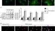
Cardiac fibrosis is a commonly seen pathophysiological process in various cardiovascular disorders, such as coronary heart disorder, hypertension, and cardiomyopathy. Cardiac fibroblast trans-differentiation into myofibroblasts (MFs) is a key link in myocardial fibrosis. LncRNA PVT1 participates in fibrotic diseases in multiple organs; however, its role and mechanism in cardiac fibrosis remain largely unknown. Human cardiac fibroblasts (HCFs) were stimulated with TGF-β1 to induce myofibroblast; Immunofluorescent staining, Immunoblotting, and fluorescence in situ hybridization were used to detect the myofibroblasts phenotypes and lnc PVT1 expression. Cell biological phenotypes induced by lnc PVT1 knockdown or overexpression were detected by CCK-8, flow cytometry, and Immunoblotting. A mouse model of myocardial fibrosis was induced using isoproterenol (ISO), and the cardiac functions were examined by echocardiography measurements, cardiac tissues by H&E, and Masson trichrome staining. In this study, TGF-β1 induced HCF transformation into myofibroblasts, as manifested as significantly increased levels of α-SMA, vimentin, collagen I, and collagen III; the expression level of lnc PVT1 expression showed to be significantly increased by TGF-β1 stimulation. The protein levels of TGF-β1, TGFBR1, and TGFBR2 were also decreased by lnc PVT1 knockdown. Under TGF-β1 stimulation, lnc PVT1 knockdown decreased FN1, α-SMA, collagen I, and collagen III protein contents, inhibited HCF cell viability and enhanced cell apoptosis, and inhibited Smad2/3 phosphorylation. Lnc PVT1 positively regulated MYC expression with or without TGF-β1 stimulation; MYC overexpression in TGF-β1-stimulated HCFs significantly attenuated the effects of lnc PVT1 knockdown on HCF proliferation and trans-differentiation to MFs. In the ISO-induced myocardial fibrosis model, lnc PVT1 knockdown partially reduced fibrotic area, improved cardiac functions, and decreased the levels of fibrotic markers. In addition, lnc PVT1 knockdown decreased MYC and CDK4 levels but increased E-cadherin in mice heart tissues. lnc PVT1 is up-regulated in cardiac fibrosis and TGF-β1-stimulated HCFs. Lnc PVT1 knockdown partially ameliorates TGF-β1-induced HCF activation and trans-differentiation into MFs in vitro and ISO-induced myocardial fibrosis in vivo, potentially through interacting with MYC and up-regulating MYC.
期刊介绍:
Molecular and Cellular Biochemistry: An International Journal for Chemical Biology in Health and Disease publishes original research papers and short communications in all areas of the biochemical sciences, emphasizing novel findings relevant to the biochemical basis of cellular function and disease processes, as well as the mechanics of action of hormones and chemical agents. Coverage includes membrane transport, receptor mechanism, immune response, secretory processes, and cytoskeletal function, as well as biochemical structure-function relationships in the cell.
In addition to the reports of original research, the journal publishes state of the art reviews. Specific subjects covered by Molecular and Cellular Biochemistry include cellular metabolism, cellular pathophysiology, enzymology, ion transport, lipid biochemistry, membrane biochemistry, molecular biology, nuclear structure and function, and protein chemistry.
 求助内容:
求助内容: 应助结果提醒方式:


