Priscilla Lamendola, Nello Cambise, Antonio Di Renzo, Lorenzo Tinti, Antonio De Vita, Saverio Tremamunno, Paola Pastena, Antonietta Belmusto, Rocco Montone, Riccardo Rinaldi, Angelo Villano, Gaetano A Lanza
{"title":"通过超声心动图运动负荷试验评估心肌桥的缺血效应","authors":"Priscilla Lamendola, Nello Cambise, Antonio Di Renzo, Lorenzo Tinti, Antonio De Vita, Saverio Tremamunno, Paola Pastena, Antonietta Belmusto, Rocco Montone, Riccardo Rinaldi, Angelo Villano, Gaetano A Lanza","doi":"10.15420/ecr.2024.03","DOIUrl":null,"url":null,"abstract":"<p><strong>Background: </strong>Detection of myocardial bridge (MB) at angiography suggests it has a role in ischaemic-related symptoms in patients with angina without obstructive coronary artery disease. However, evidence that MB may cause myocardial ischaemia is limited.</p><p><strong>Methods: </strong>We studied 41 patients with MB of the left anterior descending coronary artery and otherwise normal coronary arteries. Fourteen patients with normal coronary arteries and without MB served as controls. All subjects underwent a maximal treadmill exercise stress test (EST) under ECG monitoring. Standard and speckle-tracking echocardiography were performed at baseline and immediately after peak EST.</p><p><strong>Results: </strong>EST duration and peak heart rate and systolic pressure were similar in the two groups. A positive EST (ST-segment depression .1 mm) was found in 18 patients in the MB group (43.9%) and none in the control group (p=0.001). No abnormalities in both left ventricle systolic and diastolic function were found between the two groups in the standard echocardiographic evaluation. Global and segmental (anterior, inferior) longitudinal strain (LS) did not differ at baseline between the groups. There was a small increase in global LS during EST in MB patients but not in the control group (p=0.01). Similar trends were found for regional LSs, with differences being significant for the medium (p=0.028) and apical (p=0.032) anterior segments. No differences in echocardiographic parameters and both global and segmental LSs were observed between MB patients with ischaemic ECG changes during EST versus those without.</p><p><strong>Conclusion: </strong>Our findings do not support the notion that MB results in significant degrees of myocardial ischaemia during maximal myocardial work.</p>","PeriodicalId":93994,"journal":{"name":"European cardiology","volume":"19 ","pages":"e09"},"PeriodicalIF":0.0000,"publicationDate":"2024-06-19","publicationTypes":"Journal Article","fieldsOfStudy":null,"isOpenAccess":false,"openAccessPdf":"https://www.ncbi.nlm.nih.gov/pmc/articles/PMC11231813/pdf/","citationCount":"0","resultStr":"{\"title\":\"Assessment of the Ischaemic Effects of Myocardial Bridge by Echocardiographic Exercise Stress Test.\",\"authors\":\"Priscilla Lamendola, Nello Cambise, Antonio Di Renzo, Lorenzo Tinti, Antonio De Vita, Saverio Tremamunno, Paola Pastena, Antonietta Belmusto, Rocco Montone, Riccardo Rinaldi, Angelo Villano, Gaetano A Lanza\",\"doi\":\"10.15420/ecr.2024.03\",\"DOIUrl\":null,\"url\":null,\"abstract\":\"<p><strong>Background: </strong>Detection of myocardial bridge (MB) at angiography suggests it has a role in ischaemic-related symptoms in patients with angina without obstructive coronary artery disease. However, evidence that MB may cause myocardial ischaemia is limited.</p><p><strong>Methods: </strong>We studied 41 patients with MB of the left anterior descending coronary artery and otherwise normal coronary arteries. Fourteen patients with normal coronary arteries and without MB served as controls. All subjects underwent a maximal treadmill exercise stress test (EST) under ECG monitoring. Standard and speckle-tracking echocardiography were performed at baseline and immediately after peak EST.</p><p><strong>Results: </strong>EST duration and peak heart rate and systolic pressure were similar in the two groups. A positive EST (ST-segment depression .1 mm) was found in 18 patients in the MB group (43.9%) and none in the control group (p=0.001). No abnormalities in both left ventricle systolic and diastolic function were found between the two groups in the standard echocardiographic evaluation. Global and segmental (anterior, inferior) longitudinal strain (LS) did not differ at baseline between the groups. There was a small increase in global LS during EST in MB patients but not in the control group (p=0.01). Similar trends were found for regional LSs, with differences being significant for the medium (p=0.028) and apical (p=0.032) anterior segments. No differences in echocardiographic parameters and both global and segmental LSs were observed between MB patients with ischaemic ECG changes during EST versus those without.</p><p><strong>Conclusion: </strong>Our findings do not support the notion that MB results in significant degrees of myocardial ischaemia during maximal myocardial work.</p>\",\"PeriodicalId\":93994,\"journal\":{\"name\":\"European cardiology\",\"volume\":\"19 \",\"pages\":\"e09\"},\"PeriodicalIF\":0.0000,\"publicationDate\":\"2024-06-19\",\"publicationTypes\":\"Journal Article\",\"fieldsOfStudy\":null,\"isOpenAccess\":false,\"openAccessPdf\":\"https://www.ncbi.nlm.nih.gov/pmc/articles/PMC11231813/pdf/\",\"citationCount\":\"0\",\"resultStr\":null,\"platform\":\"Semanticscholar\",\"paperid\":null,\"PeriodicalName\":\"European cardiology\",\"FirstCategoryId\":\"1085\",\"ListUrlMain\":\"https://doi.org/10.15420/ecr.2024.03\",\"RegionNum\":0,\"RegionCategory\":null,\"ArticlePicture\":[],\"TitleCN\":null,\"AbstractTextCN\":null,\"PMCID\":null,\"EPubDate\":\"2024/1/1 0:00:00\",\"PubModel\":\"eCollection\",\"JCR\":\"\",\"JCRName\":\"\",\"Score\":null,\"Total\":0}","platform":"Semanticscholar","paperid":null,"PeriodicalName":"European cardiology","FirstCategoryId":"1085","ListUrlMain":"https://doi.org/10.15420/ecr.2024.03","RegionNum":0,"RegionCategory":null,"ArticlePicture":[],"TitleCN":null,"AbstractTextCN":null,"PMCID":null,"EPubDate":"2024/1/1 0:00:00","PubModel":"eCollection","JCR":"","JCRName":"","Score":null,"Total":0}
引用次数: 0
摘要
背景:血管造影术中发现的心肌桥(MB)表明,它在无阻塞性冠状动脉疾病的心绞痛患者出现缺血相关症状时起作用。然而,MB 可能导致心肌缺血的证据却很有限:我们研究了 41 名左前降支冠状动脉 MB 患者和其他冠状动脉正常的患者。14 名冠状动脉正常但无 MB 的患者作为对照组。所有受试者都在心电图监测下接受了最大跑步机运动负荷试验(EST)。在基线和EST峰值后立即进行标准和斑点追踪超声心动图检查:结果:两组的EST持续时间、峰值心率和收缩压相似。MB 组有 18 名患者(43.9%)发现 EST 阳性(ST 段压低 0.1 毫米),而对照组没有发现(P=0.001)。在标准超声心动图评估中,两组患者的左心室收缩和舒张功能均未发现异常。两组的整体和节段(前部、下部)纵向应变(LS)在基线时没有差异。在 EST 期间,MB 患者的整体纵向应变略有增加,而对照组则没有(P=0.01)。区域 LS 也有类似的趋势,中段(p=0.028)和心尖前段(p=0.032)的差异显著。在 EST 期间出现缺血性心电图变化的 MB 患者与未出现缺血性心电图变化的 MB 患者之间,未观察到超声心动图参数以及整体和节段 LS 的差异:我们的研究结果并不支持 MB 会在最大心肌工作期间导致严重心肌缺血的观点。
Assessment of the Ischaemic Effects of Myocardial Bridge by Echocardiographic Exercise Stress Test.
Background: Detection of myocardial bridge (MB) at angiography suggests it has a role in ischaemic-related symptoms in patients with angina without obstructive coronary artery disease. However, evidence that MB may cause myocardial ischaemia is limited.
Methods: We studied 41 patients with MB of the left anterior descending coronary artery and otherwise normal coronary arteries. Fourteen patients with normal coronary arteries and without MB served as controls. All subjects underwent a maximal treadmill exercise stress test (EST) under ECG monitoring. Standard and speckle-tracking echocardiography were performed at baseline and immediately after peak EST.
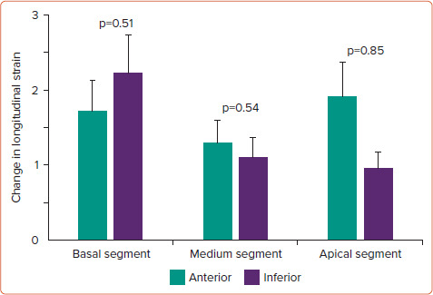
Results: EST duration and peak heart rate and systolic pressure were similar in the two groups. A positive EST (ST-segment depression .1 mm) was found in 18 patients in the MB group (43.9%) and none in the control group (p=0.001). No abnormalities in both left ventricle systolic and diastolic function were found between the two groups in the standard echocardiographic evaluation. Global and segmental (anterior, inferior) longitudinal strain (LS) did not differ at baseline between the groups. There was a small increase in global LS during EST in MB patients but not in the control group (p=0.01). Similar trends were found for regional LSs, with differences being significant for the medium (p=0.028) and apical (p=0.032) anterior segments. No differences in echocardiographic parameters and both global and segmental LSs were observed between MB patients with ischaemic ECG changes during EST versus those without.
Conclusion: Our findings do not support the notion that MB results in significant degrees of myocardial ischaemia during maximal myocardial work.

 求助内容:
求助内容: 应助结果提醒方式:


