Amr Mounir, Engy Mohamed Mostafa, Ibrahim Amer, Ahmed Abdelaleem Abdelgbar, Hamdy Osman Osman, Mostafa Abdelrahman Ahmed, Hossam Ziada, Abdel Ghany Ali El Gabbar, Mohamed Alsadawy Hassan, Alaa Mahmoud
{"title":"飞秒激光辅助角膜内环节段植入术后角膜密度的变化。","authors":"Amr Mounir, Engy Mohamed Mostafa, Ibrahim Amer, Ahmed Abdelaleem Abdelgbar, Hamdy Osman Osman, Mostafa Abdelrahman Ahmed, Hossam Ziada, Abdel Ghany Ali El Gabbar, Mohamed Alsadawy Hassan, Alaa Mahmoud","doi":"10.51329/mehdiophthal1491","DOIUrl":null,"url":null,"abstract":"<p><strong>Background: </strong>Intrastromal corneal ring segments are commonly implanted in the corneas of eyes with mild-to-moderate keratoconus; however, changes in corneal densitometry (CD) after implantation are a matter of debate in the current literature. We evaluated the changes in CD 1 and 3 months after femtosecond laser-assisted Keraring implantation.</p><p><strong>Methods: </strong>This retrospective, non-comparative, multicenter, case series study included patients with keratoconus who underwent femtosecond laser-assisted implantation of double segments with 90° and 160° arc lengths or two 160° arc length Keraring segments. Demographic and baseline clinical ophthalmic data were recorded. Corneal topography and tomography data acquired using a Pentacam HR Scheimpflug tomography system (Pentacam High Resolution; Oculus, Wetzlar, Germany) with a best-fit sphere were used as a reference surface. Using the Pentacam HR, CD measurements were acquired over a corneal area of 12 mm in total and at four concentric zones (0-2, 2-6, 6-10, and 10-12 mm) of three corneal stromal depths: 120 μm of the anterior corneal stromal layer, 60 μm of the posterior corneal stromal layer, and the central layer of stroma lying between these two layers.</p><p><strong>Results: </strong>We included 40 eyes of 40 patients, including 8 (20%) male and 32 (80%) female individuals, with a mean (standard deviation) age of 21.0 (6.4) years. We observed a significant improvement in the topographic values of steep keratometry (K), flat K, maximum K, and corneal astigmatism (all P < 0.05), but not in the mean K, thinnest corneal pachymetry, corneal thickness at the apex, back elevation, or front elevation (all P > 0.05). The mean total anterior, central, and posterior CD differed significantly among the time points, with a significant increase from the preoperative to the 1-month and 3-month postoperative visits (all P < 0.05) and no difference between those of the 1-month and 3-month postoperative visits (all P > 0.05). The mean CD for the anterior layer in the central, paracentral, and mid-peripheral zones, and the central layer in all four zones, differed significantly among time points, with a significant increase from the preoperative to the 1-month and 3-month postoperative visits (all P < 0.05), which remained unchanged from the 1-month to the 3-month postoperative visit (all P < 0.05), except for the central 2-6-mm zone, which decreased significantly from the 1-month to the 3-month postoperative visit (P < 0.001). The CD of the central 10-12-mm zone did not differ significantly in each pairwise comparison (all P > 0.05). In contrast, CD for the posterior layer in the paracentral zone decreased significantly from the preoperative to the 1-month and 3-month postoperative visits but increased, to a lesser extent, from the 1-month to the 3-month postoperative visit (all P < 0.05).</p><p><strong>Conclusions: </strong>Femtosecond laser-assisted Keraring implantation significantly changes CD, with improvement in most topography parameters. Further longitudinal studies with larger sample sizes are required to verify these preliminary findings.</p>","PeriodicalId":36524,"journal":{"name":"Medical Hypothesis, Discovery, and Innovation in Ophthalmology","volume":"13 1","pages":"27-34"},"PeriodicalIF":0.0000,"publicationDate":"2024-07-01","publicationTypes":"Journal Article","fieldsOfStudy":null,"isOpenAccess":false,"openAccessPdf":"https://www.ncbi.nlm.nih.gov/pmc/articles/PMC11227663/pdf/","citationCount":"0","resultStr":"{\"title\":\"Corneal densitometry changes after femtosecond laser-assisted intracorneal ring segments implantation in keratoconus.\",\"authors\":\"Amr Mounir, Engy Mohamed Mostafa, Ibrahim Amer, Ahmed Abdelaleem Abdelgbar, Hamdy Osman Osman, Mostafa Abdelrahman Ahmed, Hossam Ziada, Abdel Ghany Ali El Gabbar, Mohamed Alsadawy Hassan, Alaa Mahmoud\",\"doi\":\"10.51329/mehdiophthal1491\",\"DOIUrl\":null,\"url\":null,\"abstract\":\"<p><strong>Background: </strong>Intrastromal corneal ring segments are commonly implanted in the corneas of eyes with mild-to-moderate keratoconus; however, changes in corneal densitometry (CD) after implantation are a matter of debate in the current literature. We evaluated the changes in CD 1 and 3 months after femtosecond laser-assisted Keraring implantation.</p><p><strong>Methods: </strong>This retrospective, non-comparative, multicenter, case series study included patients with keratoconus who underwent femtosecond laser-assisted implantation of double segments with 90° and 160° arc lengths or two 160° arc length Keraring segments. Demographic and baseline clinical ophthalmic data were recorded. Corneal topography and tomography data acquired using a Pentacam HR Scheimpflug tomography system (Pentacam High Resolution; Oculus, Wetzlar, Germany) with a best-fit sphere were used as a reference surface. Using the Pentacam HR, CD measurements were acquired over a corneal area of 12 mm in total and at four concentric zones (0-2, 2-6, 6-10, and 10-12 mm) of three corneal stromal depths: 120 μm of the anterior corneal stromal layer, 60 μm of the posterior corneal stromal layer, and the central layer of stroma lying between these two layers.</p><p><strong>Results: </strong>We included 40 eyes of 40 patients, including 8 (20%) male and 32 (80%) female individuals, with a mean (standard deviation) age of 21.0 (6.4) years. We observed a significant improvement in the topographic values of steep keratometry (K), flat K, maximum K, and corneal astigmatism (all P < 0.05), but not in the mean K, thinnest corneal pachymetry, corneal thickness at the apex, back elevation, or front elevation (all P > 0.05). The mean total anterior, central, and posterior CD differed significantly among the time points, with a significant increase from the preoperative to the 1-month and 3-month postoperative visits (all P < 0.05) and no difference between those of the 1-month and 3-month postoperative visits (all P > 0.05). The mean CD for the anterior layer in the central, paracentral, and mid-peripheral zones, and the central layer in all four zones, differed significantly among time points, with a significant increase from the preoperative to the 1-month and 3-month postoperative visits (all P < 0.05), which remained unchanged from the 1-month to the 3-month postoperative visit (all P < 0.05), except for the central 2-6-mm zone, which decreased significantly from the 1-month to the 3-month postoperative visit (P < 0.001). The CD of the central 10-12-mm zone did not differ significantly in each pairwise comparison (all P > 0.05). In contrast, CD for the posterior layer in the paracentral zone decreased significantly from the preoperative to the 1-month and 3-month postoperative visits but increased, to a lesser extent, from the 1-month to the 3-month postoperative visit (all P < 0.05).</p><p><strong>Conclusions: </strong>Femtosecond laser-assisted Keraring implantation significantly changes CD, with improvement in most topography parameters. Further longitudinal studies with larger sample sizes are required to verify these preliminary findings.</p>\",\"PeriodicalId\":36524,\"journal\":{\"name\":\"Medical Hypothesis, Discovery, and Innovation in Ophthalmology\",\"volume\":\"13 1\",\"pages\":\"27-34\"},\"PeriodicalIF\":0.0000,\"publicationDate\":\"2024-07-01\",\"publicationTypes\":\"Journal Article\",\"fieldsOfStudy\":null,\"isOpenAccess\":false,\"openAccessPdf\":\"https://www.ncbi.nlm.nih.gov/pmc/articles/PMC11227663/pdf/\",\"citationCount\":\"0\",\"resultStr\":null,\"platform\":\"Semanticscholar\",\"paperid\":null,\"PeriodicalName\":\"Medical Hypothesis, Discovery, and Innovation in Ophthalmology\",\"FirstCategoryId\":\"1085\",\"ListUrlMain\":\"https://doi.org/10.51329/mehdiophthal1491\",\"RegionNum\":0,\"RegionCategory\":null,\"ArticlePicture\":[],\"TitleCN\":null,\"AbstractTextCN\":null,\"PMCID\":null,\"EPubDate\":\"2024/1/1 0:00:00\",\"PubModel\":\"eCollection\",\"JCR\":\"Q2\",\"JCRName\":\"Medicine\",\"Score\":null,\"Total\":0}","platform":"Semanticscholar","paperid":null,"PeriodicalName":"Medical Hypothesis, Discovery, and Innovation in Ophthalmology","FirstCategoryId":"1085","ListUrlMain":"https://doi.org/10.51329/mehdiophthal1491","RegionNum":0,"RegionCategory":null,"ArticlePicture":[],"TitleCN":null,"AbstractTextCN":null,"PMCID":null,"EPubDate":"2024/1/1 0:00:00","PubModel":"eCollection","JCR":"Q2","JCRName":"Medicine","Score":null,"Total":0}
引用次数: 0
摘要
背景:在轻度至中度角膜炎患者的角膜上通常会植入基质内角膜环段;然而,植入后角膜密度(CD)的变化在目前的文献中还存在争议。我们评估了飞秒激光辅助角膜塑形镜植入术后 1 个月和 3 个月 CD 的变化:这项回顾性、非比较性、多中心、病例系列研究纳入了在飞秒激光辅助下植入 90° 和 160° 弧长双节段或两个 160° 弧长 Keraring 节段的角膜炎患者。记录了人口统计学和眼科临床基线数据。使用 Pentacam HR Scheimpflug 层析成像系统(Pentacam 高分辨率;Oculus,德国,Wetzlar)采集的角膜地形图和层析成像数据以最佳拟合球面为参考面。使用 Pentacam HR,在总长度为 12 毫米的角膜区域和三个角膜基质深度的四个同心区(0-2、2-6、6-10 和 10-12 毫米)进行了 CD 测量:角膜前基质层 120 微米,角膜后基质层 60 微米,以及位于这两层之间的中央基质层:我们共纳入了 40 名患者的 40 只眼睛,其中男性 8 名(占 20%),女性 32 名(占 80%),平均年龄(标准差)为 21.0(6.4)岁。我们观察到,陡角膜度数(K)、平角膜度数、最大角膜度数和角膜散光的地形学值均有明显改善(P 均小于 0.05),但平均角膜度数、最薄角膜厚度、角膜顶点厚度、后方抬高和前方抬高的地形学值均无明显改善(P 均大于 0.05)。术前、术后 1 个月和 3 个月的前层、中央层和后层平均总 CD 值在不同时间点之间差异显著,术前显著增加(所有 P 均小于 0.05),术后 1 个月和 3 个月没有差异(所有 P 均大于 0.05)。中央区、旁中心区和中外周区前层以及所有四个区中央层的平均 CD 在不同时间点有显著差异,术前至术后 1 个月和 3 个月的平均 CD 显著增加(均 P < 0.05),从术后 1 个月到术后 3 个月就诊时保持不变(均 P <0.05),只有中央 2-6 mm 区从术后 1 个月到术后 3 个月就诊时明显减少(P <0.001)。中央 10-12 毫米区的 CD 在每对比较中均无明显差异(均 P > 0.05)。与此相反,旁中心区后层的 CD 从术前到术后 1 个月和 3 个月时明显减少,但从术后 1 个月到术后 3 个月时增加较少(均 P <0.05):结论:飞秒激光辅助下的 Keraring 植入术能显著改变 CD,改善大多数地形参数。要验证这些初步研究结果,还需要更多样本量的纵向研究。
Corneal densitometry changes after femtosecond laser-assisted intracorneal ring segments implantation in keratoconus.
Background: Intrastromal corneal ring segments are commonly implanted in the corneas of eyes with mild-to-moderate keratoconus; however, changes in corneal densitometry (CD) after implantation are a matter of debate in the current literature. We evaluated the changes in CD 1 and 3 months after femtosecond laser-assisted Keraring implantation.
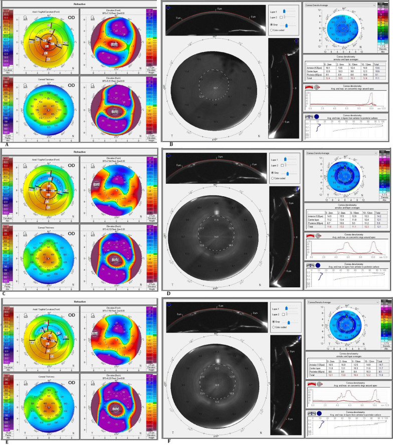
Methods: This retrospective, non-comparative, multicenter, case series study included patients with keratoconus who underwent femtosecond laser-assisted implantation of double segments with 90° and 160° arc lengths or two 160° arc length Keraring segments. Demographic and baseline clinical ophthalmic data were recorded. Corneal topography and tomography data acquired using a Pentacam HR Scheimpflug tomography system (Pentacam High Resolution; Oculus, Wetzlar, Germany) with a best-fit sphere were used as a reference surface. Using the Pentacam HR, CD measurements were acquired over a corneal area of 12 mm in total and at four concentric zones (0-2, 2-6, 6-10, and 10-12 mm) of three corneal stromal depths: 120 μm of the anterior corneal stromal layer, 60 μm of the posterior corneal stromal layer, and the central layer of stroma lying between these two layers.
Results: We included 40 eyes of 40 patients, including 8 (20%) male and 32 (80%) female individuals, with a mean (standard deviation) age of 21.0 (6.4) years. We observed a significant improvement in the topographic values of steep keratometry (K), flat K, maximum K, and corneal astigmatism (all P < 0.05), but not in the mean K, thinnest corneal pachymetry, corneal thickness at the apex, back elevation, or front elevation (all P > 0.05). The mean total anterior, central, and posterior CD differed significantly among the time points, with a significant increase from the preoperative to the 1-month and 3-month postoperative visits (all P < 0.05) and no difference between those of the 1-month and 3-month postoperative visits (all P > 0.05). The mean CD for the anterior layer in the central, paracentral, and mid-peripheral zones, and the central layer in all four zones, differed significantly among time points, with a significant increase from the preoperative to the 1-month and 3-month postoperative visits (all P < 0.05), which remained unchanged from the 1-month to the 3-month postoperative visit (all P < 0.05), except for the central 2-6-mm zone, which decreased significantly from the 1-month to the 3-month postoperative visit (P < 0.001). The CD of the central 10-12-mm zone did not differ significantly in each pairwise comparison (all P > 0.05). In contrast, CD for the posterior layer in the paracentral zone decreased significantly from the preoperative to the 1-month and 3-month postoperative visits but increased, to a lesser extent, from the 1-month to the 3-month postoperative visit (all P < 0.05).
Conclusions: Femtosecond laser-assisted Keraring implantation significantly changes CD, with improvement in most topography parameters. Further longitudinal studies with larger sample sizes are required to verify these preliminary findings.

 求助内容:
求助内容: 应助结果提醒方式:


