{"title":"通过基于非对比 CT 的深度学习检测血管内主动脉修复术后的内渗漏","authors":"Qingqi Yang, Jinglang Hu, Yingqi Luo, Dongdong Jia, Nuo Chen, Chen Yao, Ridong Wu","doi":"10.1007/s00270-024-03805-x","DOIUrl":null,"url":null,"abstract":"<p><strong>Objectives: </strong>To develop and validate a deep learning model for detecting post-endovascular aortic repair (EVAR) endoleak from non-contrast CT.</p><p><strong>Methods: </strong>This retrospective study involved 245 patients who underwent EVAR between September 2016 and December 2022. All patients underwent both non-enhanced and enhanced follow-up CT. The presence of endoleak was evaluated based on computed tomography angiography (CTA) and radiology reports. First, the aneurysm sac was segmented, and radiomic features were extracted on non-contrast CT. Statistical analysis was conducted to investigate differences in shape and density characteristics between aneurysm sacs with and without endoleak. Subsequently, a deep learning model was trained to generate predicted segmentation of the endoleak. A binary decision was made based on whether the model produced a segmentation to detect the presence of endoleak. The absence of a predicted segmentation indicated no endoleak, while the presence of a predicted segmentation indicated endoleak. Finally, the performance of the model was evaluated by comparing the predicted segmentation with the reference segmentation obtained from CTA. Model performance was assessed using metrics such as dice similarity coefficient, sensitivity, specificity, and the area under the curve (AUC).</p><p><strong>Results: </strong>This study finally included 85 patients with endoleak and 82 patients without endoleak. Compared to patients without endoleak, patients with endoleak had higher CT values and greater dispersion. The AUC in validation group was 0.951, dice similarity coefficient was 0.814, sensitivity was 0.877, and specificity was 0.884.</p><p><strong>Conclusion: </strong>This deep learning model based on non-contrast CT can detect endoleak after EVAR with high sensitivity.</p>","PeriodicalId":9591,"journal":{"name":"CardioVascular and Interventional Radiology","volume":null,"pages":null},"PeriodicalIF":2.8000,"publicationDate":"2024-09-01","publicationTypes":"Journal Article","fieldsOfStudy":null,"isOpenAccess":false,"openAccessPdf":"","citationCount":"0","resultStr":"{\"title\":\"Detection of Endoleak after Endovascular Aortic Repair through Deep Learning Based on Non-contrast CT.\",\"authors\":\"Qingqi Yang, Jinglang Hu, Yingqi Luo, Dongdong Jia, Nuo Chen, Chen Yao, Ridong Wu\",\"doi\":\"10.1007/s00270-024-03805-x\",\"DOIUrl\":null,\"url\":null,\"abstract\":\"<p><strong>Objectives: </strong>To develop and validate a deep learning model for detecting post-endovascular aortic repair (EVAR) endoleak from non-contrast CT.</p><p><strong>Methods: </strong>This retrospective study involved 245 patients who underwent EVAR between September 2016 and December 2022. All patients underwent both non-enhanced and enhanced follow-up CT. The presence of endoleak was evaluated based on computed tomography angiography (CTA) and radiology reports. First, the aneurysm sac was segmented, and radiomic features were extracted on non-contrast CT. Statistical analysis was conducted to investigate differences in shape and density characteristics between aneurysm sacs with and without endoleak. Subsequently, a deep learning model was trained to generate predicted segmentation of the endoleak. A binary decision was made based on whether the model produced a segmentation to detect the presence of endoleak. The absence of a predicted segmentation indicated no endoleak, while the presence of a predicted segmentation indicated endoleak. Finally, the performance of the model was evaluated by comparing the predicted segmentation with the reference segmentation obtained from CTA. Model performance was assessed using metrics such as dice similarity coefficient, sensitivity, specificity, and the area under the curve (AUC).</p><p><strong>Results: </strong>This study finally included 85 patients with endoleak and 82 patients without endoleak. Compared to patients without endoleak, patients with endoleak had higher CT values and greater dispersion. The AUC in validation group was 0.951, dice similarity coefficient was 0.814, sensitivity was 0.877, and specificity was 0.884.</p><p><strong>Conclusion: </strong>This deep learning model based on non-contrast CT can detect endoleak after EVAR with high sensitivity.</p>\",\"PeriodicalId\":9591,\"journal\":{\"name\":\"CardioVascular and Interventional Radiology\",\"volume\":null,\"pages\":null},\"PeriodicalIF\":2.8000,\"publicationDate\":\"2024-09-01\",\"publicationTypes\":\"Journal Article\",\"fieldsOfStudy\":null,\"isOpenAccess\":false,\"openAccessPdf\":\"\",\"citationCount\":\"0\",\"resultStr\":null,\"platform\":\"Semanticscholar\",\"paperid\":null,\"PeriodicalName\":\"CardioVascular and Interventional Radiology\",\"FirstCategoryId\":\"3\",\"ListUrlMain\":\"https://doi.org/10.1007/s00270-024-03805-x\",\"RegionNum\":3,\"RegionCategory\":\"医学\",\"ArticlePicture\":[],\"TitleCN\":null,\"AbstractTextCN\":null,\"PMCID\":null,\"EPubDate\":\"2024/7/8 0:00:00\",\"PubModel\":\"Epub\",\"JCR\":\"Q2\",\"JCRName\":\"CARDIAC & CARDIOVASCULAR SYSTEMS\",\"Score\":null,\"Total\":0}","platform":"Semanticscholar","paperid":null,"PeriodicalName":"CardioVascular and Interventional Radiology","FirstCategoryId":"3","ListUrlMain":"https://doi.org/10.1007/s00270-024-03805-x","RegionNum":3,"RegionCategory":"医学","ArticlePicture":[],"TitleCN":null,"AbstractTextCN":null,"PMCID":null,"EPubDate":"2024/7/8 0:00:00","PubModel":"Epub","JCR":"Q2","JCRName":"CARDIAC & CARDIOVASCULAR SYSTEMS","Score":null,"Total":0}
Detection of Endoleak after Endovascular Aortic Repair through Deep Learning Based on Non-contrast CT.
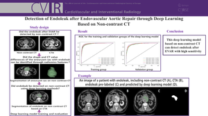
Objectives: To develop and validate a deep learning model for detecting post-endovascular aortic repair (EVAR) endoleak from non-contrast CT.
Methods: This retrospective study involved 245 patients who underwent EVAR between September 2016 and December 2022. All patients underwent both non-enhanced and enhanced follow-up CT. The presence of endoleak was evaluated based on computed tomography angiography (CTA) and radiology reports. First, the aneurysm sac was segmented, and radiomic features were extracted on non-contrast CT. Statistical analysis was conducted to investigate differences in shape and density characteristics between aneurysm sacs with and without endoleak. Subsequently, a deep learning model was trained to generate predicted segmentation of the endoleak. A binary decision was made based on whether the model produced a segmentation to detect the presence of endoleak. The absence of a predicted segmentation indicated no endoleak, while the presence of a predicted segmentation indicated endoleak. Finally, the performance of the model was evaluated by comparing the predicted segmentation with the reference segmentation obtained from CTA. Model performance was assessed using metrics such as dice similarity coefficient, sensitivity, specificity, and the area under the curve (AUC).
Results: This study finally included 85 patients with endoleak and 82 patients without endoleak. Compared to patients without endoleak, patients with endoleak had higher CT values and greater dispersion. The AUC in validation group was 0.951, dice similarity coefficient was 0.814, sensitivity was 0.877, and specificity was 0.884.
Conclusion: This deep learning model based on non-contrast CT can detect endoleak after EVAR with high sensitivity.
期刊介绍:
CardioVascular and Interventional Radiology (CVIR) is the official journal of the Cardiovascular and Interventional Radiological Society of Europe, and is also the official organ of a number of additional distinguished national and international interventional radiological societies. CVIR publishes double blinded peer-reviewed original research work including clinical and laboratory investigations, technical notes, case reports, works in progress, and letters to the editor, as well as review articles, pictorial essays, editorials, and special invited submissions in the field of vascular and interventional radiology. Beside the communication of the latest research results in this field, it is also the aim of CVIR to support continuous medical education. Articles that are accepted for publication are done so with the understanding that they, or their substantive contents, have not been and will not be submitted to any other publication.
 求助内容:
求助内容: 应助结果提醒方式:


