Odianosen J. Eigbire-Molen , Clarissa A. Cassol , Daniel J. Kenan , Johnathan O.H. Napier , Lyle J. Burdine , Shana M. Coley , Shree G. Sharma
{"title":"基于智能手机的机器学习模型用于医学肾活检的实时评估","authors":"Odianosen J. Eigbire-Molen , Clarissa A. Cassol , Daniel J. Kenan , Johnathan O.H. Napier , Lyle J. Burdine , Shana M. Coley , Shree G. Sharma","doi":"10.1016/j.jpi.2024.100385","DOIUrl":null,"url":null,"abstract":"<div><h3>Background</h3><p>Kidney biopsy is the gold-standard for diagnosing medical renal diseases, but the accuracy of the diagnosis greatly depends on the quality of the biopsy specimen, particularly the amount of renal cortex obtained. Inadequate biopsies, characterized by insufficient cortex or predominant medulla, can lead to inconclusive or incorrect diagnoses, and repeat biopsy. Unfortunately, there has been a concerning increase in the rate of inadequate kidney biopsies, and not all medical centers have access to trained professionals who can assess biopsy adequacy in real time. In response to this challenge, we aimed to develop a machine learning model capable of assessing the percentage cortex of each biopsy pass using smartphone images of the kidney biopsy tissue at the time of biopsy.</p></div><div><h3>Methods</h3><p>747 kidney biopsy cores and corresponding smartphone macro images were collected from five unused deceased donor kidneys. Each core was imaged, formalin-fixed, sectioned, and stained with Periodic acid–Schiff (PAS) to determine cortex percentage. The fresh unfixed core images were captured using the macro camera on an iPhone 13 Pro. Two experienced renal pathologists independently reviewed the PAS-stained sections to determine the cortex percentage. For the purpose of this study, the biopsies with less than 30% cortex were labeled as inadequate, while those with 30% or more cortex were classified as adequate. The dataset was divided into training (<em>n</em>=643), validation (<em>n</em>=30), and test (<em>n</em>=74) sets. Preprocessing steps involved converting High-Efficiency Image Container iPhone format images to JPEG, normalization, and renal tissue segmentation using a U-Net deep learning model. Subsequently, a classification deep learning model was trained on the renal tissue region of interest and corresponding class label.</p></div><div><h3>Results</h3><p>The deep learning model achieved an accuracy of 85% on the training data. On the independent test dataset, the model exhibited an accuracy of 81%. For inadequate samples in the test dataset, the model showed a sensitivity of 71%, suggesting its capability to identify cases with inadequate cortical representation. The area under the receiver-operating curve (AUC-ROC) on the test dataset was 0.80.</p></div><div><h3>Conclusion</h3><p>We successfully developed and tested a machine learning model for classifying smartphone images of kidney biopsies as either adequate or inadequate, based on the amount of cortex determined by expert renal pathologists. The model's promising results suggest its potential as a smartphone application to assist real-time assessment of kidney biopsy tissue, particularly in settings with limited access to trained personnel. Further refinements and validations are warranted to optimize the model's performance.</p></div>","PeriodicalId":37769,"journal":{"name":"Journal of Pathology Informatics","volume":"15 ","pages":"Article 100385"},"PeriodicalIF":0.0000,"publicationDate":"2024-05-31","publicationTypes":"Journal Article","fieldsOfStudy":null,"isOpenAccess":false,"openAccessPdf":"https://www.sciencedirect.com/science/article/pii/S2153353924000245/pdfft?md5=aa0cdf6fbf647b60d197599f7a7fc32d&pid=1-s2.0-S2153353924000245-main.pdf","citationCount":"0","resultStr":"{\"title\":\"Smartphone-based machine learning model for real-time assessment of medical kidney biopsy\",\"authors\":\"Odianosen J. Eigbire-Molen , Clarissa A. Cassol , Daniel J. Kenan , Johnathan O.H. Napier , Lyle J. Burdine , Shana M. Coley , Shree G. Sharma\",\"doi\":\"10.1016/j.jpi.2024.100385\",\"DOIUrl\":null,\"url\":null,\"abstract\":\"<div><h3>Background</h3><p>Kidney biopsy is the gold-standard for diagnosing medical renal diseases, but the accuracy of the diagnosis greatly depends on the quality of the biopsy specimen, particularly the amount of renal cortex obtained. Inadequate biopsies, characterized by insufficient cortex or predominant medulla, can lead to inconclusive or incorrect diagnoses, and repeat biopsy. Unfortunately, there has been a concerning increase in the rate of inadequate kidney biopsies, and not all medical centers have access to trained professionals who can assess biopsy adequacy in real time. In response to this challenge, we aimed to develop a machine learning model capable of assessing the percentage cortex of each biopsy pass using smartphone images of the kidney biopsy tissue at the time of biopsy.</p></div><div><h3>Methods</h3><p>747 kidney biopsy cores and corresponding smartphone macro images were collected from five unused deceased donor kidneys. Each core was imaged, formalin-fixed, sectioned, and stained with Periodic acid–Schiff (PAS) to determine cortex percentage. The fresh unfixed core images were captured using the macro camera on an iPhone 13 Pro. Two experienced renal pathologists independently reviewed the PAS-stained sections to determine the cortex percentage. For the purpose of this study, the biopsies with less than 30% cortex were labeled as inadequate, while those with 30% or more cortex were classified as adequate. The dataset was divided into training (<em>n</em>=643), validation (<em>n</em>=30), and test (<em>n</em>=74) sets. Preprocessing steps involved converting High-Efficiency Image Container iPhone format images to JPEG, normalization, and renal tissue segmentation using a U-Net deep learning model. Subsequently, a classification deep learning model was trained on the renal tissue region of interest and corresponding class label.</p></div><div><h3>Results</h3><p>The deep learning model achieved an accuracy of 85% on the training data. On the independent test dataset, the model exhibited an accuracy of 81%. For inadequate samples in the test dataset, the model showed a sensitivity of 71%, suggesting its capability to identify cases with inadequate cortical representation. The area under the receiver-operating curve (AUC-ROC) on the test dataset was 0.80.</p></div><div><h3>Conclusion</h3><p>We successfully developed and tested a machine learning model for classifying smartphone images of kidney biopsies as either adequate or inadequate, based on the amount of cortex determined by expert renal pathologists. The model's promising results suggest its potential as a smartphone application to assist real-time assessment of kidney biopsy tissue, particularly in settings with limited access to trained personnel. Further refinements and validations are warranted to optimize the model's performance.</p></div>\",\"PeriodicalId\":37769,\"journal\":{\"name\":\"Journal of Pathology Informatics\",\"volume\":\"15 \",\"pages\":\"Article 100385\"},\"PeriodicalIF\":0.0000,\"publicationDate\":\"2024-05-31\",\"publicationTypes\":\"Journal Article\",\"fieldsOfStudy\":null,\"isOpenAccess\":false,\"openAccessPdf\":\"https://www.sciencedirect.com/science/article/pii/S2153353924000245/pdfft?md5=aa0cdf6fbf647b60d197599f7a7fc32d&pid=1-s2.0-S2153353924000245-main.pdf\",\"citationCount\":\"0\",\"resultStr\":null,\"platform\":\"Semanticscholar\",\"paperid\":null,\"PeriodicalName\":\"Journal of Pathology Informatics\",\"FirstCategoryId\":\"1085\",\"ListUrlMain\":\"https://www.sciencedirect.com/science/article/pii/S2153353924000245\",\"RegionNum\":0,\"RegionCategory\":null,\"ArticlePicture\":[],\"TitleCN\":null,\"AbstractTextCN\":null,\"PMCID\":null,\"EPubDate\":\"\",\"PubModel\":\"\",\"JCR\":\"Q2\",\"JCRName\":\"Medicine\",\"Score\":null,\"Total\":0}","platform":"Semanticscholar","paperid":null,"PeriodicalName":"Journal of Pathology Informatics","FirstCategoryId":"1085","ListUrlMain":"https://www.sciencedirect.com/science/article/pii/S2153353924000245","RegionNum":0,"RegionCategory":null,"ArticlePicture":[],"TitleCN":null,"AbstractTextCN":null,"PMCID":null,"EPubDate":"","PubModel":"","JCR":"Q2","JCRName":"Medicine","Score":null,"Total":0}
引用次数: 0
摘要
背景肾活检是诊断内科肾脏疾病的金标准,但诊断的准确性在很大程度上取决于活检标本的质量,尤其是获得的肾皮质的数量。活检不充分,表现为皮质不足或髓质占优势,可导致诊断不确定或不正确,并导致重复活检。遗憾的是,肾脏活检不充分的比例一直在上升,而且并非所有医疗中心都有训练有素的专业人员来实时评估活检是否充分。为了应对这一挑战,我们旨在开发一种机器学习模型,该模型能够利用活检时肾脏活检组织的智能手机图像评估每次活检的皮质百分比。每个肾芯都经过成像、福尔马林固定、切片和过硫酸希夫(PAS)染色,以确定皮质百分比。使用 iPhone 13 Pro 的微距摄像头拍摄了新鲜的未固定肾芯图像。两名经验丰富的肾脏病理学家独立审查 PAS 染色切片,以确定皮质百分比。在本研究中,皮质少于 30% 的活检样本被标记为皮质不足,而皮质达到或超过 30% 的活检样本则被归类为皮质充足。数据集分为训练集(样本数=643)、验证集(样本数=30)和测试集(样本数=74)。预处理步骤包括将高效图像容器 iPhone 格式图像转换为 JPEG、归一化,以及使用 U-Net 深度学习模型进行肾组织分割。随后,根据感兴趣的肾组织区域和相应的类标签训练分类深度学习模型。在独立测试数据集上,该模型的准确率为 81%。对于测试数据集中的不足样本,该模型显示出 71% 的灵敏度,表明它有能力识别皮质表征不足的病例。结论我们成功开发并测试了一种机器学习模型,该模型可根据肾脏病理专家确定的皮质数量,将肾脏活检的智能手机图像分类为充分或不充分。该模型取得了令人鼓舞的结果,表明它有潜力作为智能手机应用来协助实时评估肾活检组织,尤其是在训练有素的人员有限的情况下。为了优化该模型的性能,还需要进一步的改进和验证。
Smartphone-based machine learning model for real-time assessment of medical kidney biopsy
Background
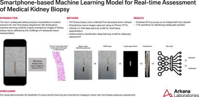
Kidney biopsy is the gold-standard for diagnosing medical renal diseases, but the accuracy of the diagnosis greatly depends on the quality of the biopsy specimen, particularly the amount of renal cortex obtained. Inadequate biopsies, characterized by insufficient cortex or predominant medulla, can lead to inconclusive or incorrect diagnoses, and repeat biopsy. Unfortunately, there has been a concerning increase in the rate of inadequate kidney biopsies, and not all medical centers have access to trained professionals who can assess biopsy adequacy in real time. In response to this challenge, we aimed to develop a machine learning model capable of assessing the percentage cortex of each biopsy pass using smartphone images of the kidney biopsy tissue at the time of biopsy.
Methods
747 kidney biopsy cores and corresponding smartphone macro images were collected from five unused deceased donor kidneys. Each core was imaged, formalin-fixed, sectioned, and stained with Periodic acid–Schiff (PAS) to determine cortex percentage. The fresh unfixed core images were captured using the macro camera on an iPhone 13 Pro. Two experienced renal pathologists independently reviewed the PAS-stained sections to determine the cortex percentage. For the purpose of this study, the biopsies with less than 30% cortex were labeled as inadequate, while those with 30% or more cortex were classified as adequate. The dataset was divided into training (n=643), validation (n=30), and test (n=74) sets. Preprocessing steps involved converting High-Efficiency Image Container iPhone format images to JPEG, normalization, and renal tissue segmentation using a U-Net deep learning model. Subsequently, a classification deep learning model was trained on the renal tissue region of interest and corresponding class label.
Results
The deep learning model achieved an accuracy of 85% on the training data. On the independent test dataset, the model exhibited an accuracy of 81%. For inadequate samples in the test dataset, the model showed a sensitivity of 71%, suggesting its capability to identify cases with inadequate cortical representation. The area under the receiver-operating curve (AUC-ROC) on the test dataset was 0.80.
Conclusion
We successfully developed and tested a machine learning model for classifying smartphone images of kidney biopsies as either adequate or inadequate, based on the amount of cortex determined by expert renal pathologists. The model's promising results suggest its potential as a smartphone application to assist real-time assessment of kidney biopsy tissue, particularly in settings with limited access to trained personnel. Further refinements and validations are warranted to optimize the model's performance.
期刊介绍:
The Journal of Pathology Informatics (JPI) is an open access peer-reviewed journal dedicated to the advancement of pathology informatics. This is the official journal of the Association for Pathology Informatics (API). The journal aims to publish broadly about pathology informatics and freely disseminate all articles worldwide. This journal is of interest to pathologists, informaticians, academics, researchers, health IT specialists, information officers, IT staff, vendors, and anyone with an interest in informatics. We encourage submissions from anyone with an interest in the field of pathology informatics. We publish all types of papers related to pathology informatics including original research articles, technical notes, reviews, viewpoints, commentaries, editorials, symposia, meeting abstracts, book reviews, and correspondence to the editors. All submissions are subject to rigorous peer review by the well-regarded editorial board and by expert referees in appropriate specialties.

 求助内容:
求助内容: 应助结果提醒方式:


