Sabine Wagner, Christian Ewald, Diana Freitag, Karl-Heinz Herrmann, Arend Koch, Johannes Bauer, Thomas J Vogl, André Kemmling, Hubert Gufler
{"title":"利用核磁共振成像预测小鼠异位胶质母细胞瘤移植成功率的放射组学和视觉分析。","authors":"Sabine Wagner, Christian Ewald, Diana Freitag, Karl-Heinz Herrmann, Arend Koch, Johannes Bauer, Thomas J Vogl, André Kemmling, Hubert Gufler","doi":"10.1007/s11060-024-04725-z","DOIUrl":null,"url":null,"abstract":"<p><strong>Background: </strong>Quantifying tumor growth and treatment response noninvasively poses a challenge to all experimental tumor models. The aim of our study was, to assess the value of quantitative and visual examination and radiomic feature analysis of high-resolution MR images of heterotopic glioblastoma xenografts in mice to determine tumor cell proliferation (TCP).</p><p><strong>Methods: </strong>Human glioblastoma cells were injected subcutaneously into both flanks of immunodeficient mice and followed up on a 3 T MR scanner. Volumes and signal intensities were calculated. Visual assessment of the internal tumor structure was based on a scoring system. Radiomic feature analysis was performed using MaZda software. The results were correlated with histopathology and immunochemistry.</p><p><strong>Results: </strong>21 tumors in 14 animals were analyzed. The volumes of xenografts with high TCP (H-TCP) increased, whereas those with low TCP (L-TCP) or no TCP (N-TCP) continued to decrease over time (p < 0.05). A low intensity rim (rim sign) on unenhanced T1-weighted images provided the highest diagnostic accuracy at visual analysis for assessing H-TCP (p < 0.05). Applying radiomic feature analysis, wavelet transform parameters were best for distinguishing between H-TCP and L-TCP / N-TCP (p < 0.05).</p><p><strong>Conclusion: </strong>Visual and radiomic feature analysis of the internal structure of heterotopically implanted glioblastomas provide reproducible and quantifiable results to predict the success of transplantation.</p>","PeriodicalId":16425,"journal":{"name":"Journal of Neuro-Oncology","volume":null,"pages":null},"PeriodicalIF":3.2000,"publicationDate":"2024-09-01","publicationTypes":"Journal Article","fieldsOfStudy":null,"isOpenAccess":false,"openAccessPdf":"https://www.ncbi.nlm.nih.gov/pmc/articles/PMC11341603/pdf/","citationCount":"0","resultStr":"{\"title\":\"Radiomics and visual analysis for predicting success of transplantation of heterotopic glioblastoma in mice with MRI.\",\"authors\":\"Sabine Wagner, Christian Ewald, Diana Freitag, Karl-Heinz Herrmann, Arend Koch, Johannes Bauer, Thomas J Vogl, André Kemmling, Hubert Gufler\",\"doi\":\"10.1007/s11060-024-04725-z\",\"DOIUrl\":null,\"url\":null,\"abstract\":\"<p><strong>Background: </strong>Quantifying tumor growth and treatment response noninvasively poses a challenge to all experimental tumor models. The aim of our study was, to assess the value of quantitative and visual examination and radiomic feature analysis of high-resolution MR images of heterotopic glioblastoma xenografts in mice to determine tumor cell proliferation (TCP).</p><p><strong>Methods: </strong>Human glioblastoma cells were injected subcutaneously into both flanks of immunodeficient mice and followed up on a 3 T MR scanner. Volumes and signal intensities were calculated. Visual assessment of the internal tumor structure was based on a scoring system. Radiomic feature analysis was performed using MaZda software. The results were correlated with histopathology and immunochemistry.</p><p><strong>Results: </strong>21 tumors in 14 animals were analyzed. The volumes of xenografts with high TCP (H-TCP) increased, whereas those with low TCP (L-TCP) or no TCP (N-TCP) continued to decrease over time (p < 0.05). A low intensity rim (rim sign) on unenhanced T1-weighted images provided the highest diagnostic accuracy at visual analysis for assessing H-TCP (p < 0.05). Applying radiomic feature analysis, wavelet transform parameters were best for distinguishing between H-TCP and L-TCP / N-TCP (p < 0.05).</p><p><strong>Conclusion: </strong>Visual and radiomic feature analysis of the internal structure of heterotopically implanted glioblastomas provide reproducible and quantifiable results to predict the success of transplantation.</p>\",\"PeriodicalId\":16425,\"journal\":{\"name\":\"Journal of Neuro-Oncology\",\"volume\":null,\"pages\":null},\"PeriodicalIF\":3.2000,\"publicationDate\":\"2024-09-01\",\"publicationTypes\":\"Journal Article\",\"fieldsOfStudy\":null,\"isOpenAccess\":false,\"openAccessPdf\":\"https://www.ncbi.nlm.nih.gov/pmc/articles/PMC11341603/pdf/\",\"citationCount\":\"0\",\"resultStr\":null,\"platform\":\"Semanticscholar\",\"paperid\":null,\"PeriodicalName\":\"Journal of Neuro-Oncology\",\"FirstCategoryId\":\"3\",\"ListUrlMain\":\"https://doi.org/10.1007/s11060-024-04725-z\",\"RegionNum\":2,\"RegionCategory\":\"医学\",\"ArticlePicture\":[],\"TitleCN\":null,\"AbstractTextCN\":null,\"PMCID\":null,\"EPubDate\":\"2024/7/3 0:00:00\",\"PubModel\":\"Epub\",\"JCR\":\"Q2\",\"JCRName\":\"CLINICAL NEUROLOGY\",\"Score\":null,\"Total\":0}","platform":"Semanticscholar","paperid":null,"PeriodicalName":"Journal of Neuro-Oncology","FirstCategoryId":"3","ListUrlMain":"https://doi.org/10.1007/s11060-024-04725-z","RegionNum":2,"RegionCategory":"医学","ArticlePicture":[],"TitleCN":null,"AbstractTextCN":null,"PMCID":null,"EPubDate":"2024/7/3 0:00:00","PubModel":"Epub","JCR":"Q2","JCRName":"CLINICAL NEUROLOGY","Score":null,"Total":0}
Radiomics and visual analysis for predicting success of transplantation of heterotopic glioblastoma in mice with MRI.
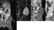
Background: Quantifying tumor growth and treatment response noninvasively poses a challenge to all experimental tumor models. The aim of our study was, to assess the value of quantitative and visual examination and radiomic feature analysis of high-resolution MR images of heterotopic glioblastoma xenografts in mice to determine tumor cell proliferation (TCP).
Methods: Human glioblastoma cells were injected subcutaneously into both flanks of immunodeficient mice and followed up on a 3 T MR scanner. Volumes and signal intensities were calculated. Visual assessment of the internal tumor structure was based on a scoring system. Radiomic feature analysis was performed using MaZda software. The results were correlated with histopathology and immunochemistry.
Results: 21 tumors in 14 animals were analyzed. The volumes of xenografts with high TCP (H-TCP) increased, whereas those with low TCP (L-TCP) or no TCP (N-TCP) continued to decrease over time (p < 0.05). A low intensity rim (rim sign) on unenhanced T1-weighted images provided the highest diagnostic accuracy at visual analysis for assessing H-TCP (p < 0.05). Applying radiomic feature analysis, wavelet transform parameters were best for distinguishing between H-TCP and L-TCP / N-TCP (p < 0.05).
Conclusion: Visual and radiomic feature analysis of the internal structure of heterotopically implanted glioblastomas provide reproducible and quantifiable results to predict the success of transplantation.
期刊介绍:
The Journal of Neuro-Oncology is a multi-disciplinary journal encompassing basic, applied, and clinical investigations in all research areas as they relate to cancer and the central nervous system. It provides a single forum for communication among neurologists, neurosurgeons, radiotherapists, medical oncologists, neuropathologists, neurodiagnosticians, and laboratory-based oncologists conducting relevant research. The Journal of Neuro-Oncology does not seek to isolate the field, but rather to focus the efforts of many disciplines in one publication through a format which pulls together these diverse interests. More than any other field of oncology, cancer of the central nervous system requires multi-disciplinary approaches. To alleviate having to scan dozens of journals of cell biology, pathology, laboratory and clinical endeavours, JNO is a periodical in which current, high-quality, relevant research in all aspects of neuro-oncology may be found.

 求助内容:
求助内容: 应助结果提醒方式:


