{"title":"成纤维细胞活化蛋白-α在人体深静脉血栓中的表达。","authors":"Nobuyuki Oguri , Toshihiro Gi , Eriko Nakamura , Eiji Furukoji , Hiroki Goto , Kazunari Maekawa , Atsushi B. Tsuji , Ryuichi Nishii , Murasaki Aman , Sayaka Moriguchi-Goto , Tatefumi Sakae , Minako Azuma , Atsushi Yamashita","doi":"10.1016/j.thromres.2024.109075","DOIUrl":null,"url":null,"abstract":"<div><h3>Background</h3><p>Fibroblast activation protein-α (FAP), a type-II transmembrane serine protease, is associated with wound healing, cancer-associated fibroblasts, and chronic fibrosing diseases. However, its expression in deep vein thrombosis (DVT) remains unclear. Therefore, this study investigated FAP expression and localization in DVT.</p></div><div><h3>Methods</h3><p>We performed pathological analyses of the aspirated thrombi of patients with DVT (<em>n</em> = 14), classifying thrombotic areas in terms of fresh, cellular lysis, and organizing reaction components. The organizing reaction included endothelialization and fibroblastic reaction. We immunohistochemically examined FAP-expressed areas and cells, and finally analyzed FAP expression in cultured dermal fibroblasts.</p></div><div><h3>Results</h3><p>All the aspirated thrombi showed a heterogeneous mixture of at least two of the three thrombotic areas. Specifically, 83 % of aspirated thrombi showed fresh and organizing reaction components. Immunohistochemical expression of FAP was restricted to the organizing area. Double immunofluorescence staining showed that FAP in the thrombi was mainly expressed in vimentin-positive or α-smooth muscle actin-positive fibroblasts. Some CD163-positive macrophages expressed FAP. FAP mRNA and protein levels were higher in fibroblasts with low-proliferative activity cultured under 0.1 % fetal bovine serum (FBS) than that under 10 % FBS. Fibroblasts cultured in 10 % FBS showed a significant decrease in <em>FAP</em> mRNA levels following supplementation with hemin, but not with thrombin.</p></div><div><h3>Conclusions</h3><p>The heterogeneous composition of venous thrombi suggests a multistep thrombus formation process in human DVT. Further, fibroblasts or myofibroblasts may express FAP during the organizing process. FAP expression may be higher in fibroblasts with low proliferative activity.</p></div>","PeriodicalId":23064,"journal":{"name":"Thrombosis research","volume":null,"pages":null},"PeriodicalIF":3.7000,"publicationDate":"2024-06-25","publicationTypes":"Journal Article","fieldsOfStudy":null,"isOpenAccess":false,"openAccessPdf":"https://www.sciencedirect.com/science/article/pii/S004938482400207X/pdfft?md5=73ac63c826d44103e3e2c69276ce5574&pid=1-s2.0-S004938482400207X-main.pdf","citationCount":"0","resultStr":"{\"title\":\"Expression of fibroblast activation protein-α in human deep vein thrombosis\",\"authors\":\"Nobuyuki Oguri , Toshihiro Gi , Eriko Nakamura , Eiji Furukoji , Hiroki Goto , Kazunari Maekawa , Atsushi B. Tsuji , Ryuichi Nishii , Murasaki Aman , Sayaka Moriguchi-Goto , Tatefumi Sakae , Minako Azuma , Atsushi Yamashita\",\"doi\":\"10.1016/j.thromres.2024.109075\",\"DOIUrl\":null,\"url\":null,\"abstract\":\"<div><h3>Background</h3><p>Fibroblast activation protein-α (FAP), a type-II transmembrane serine protease, is associated with wound healing, cancer-associated fibroblasts, and chronic fibrosing diseases. However, its expression in deep vein thrombosis (DVT) remains unclear. Therefore, this study investigated FAP expression and localization in DVT.</p></div><div><h3>Methods</h3><p>We performed pathological analyses of the aspirated thrombi of patients with DVT (<em>n</em> = 14), classifying thrombotic areas in terms of fresh, cellular lysis, and organizing reaction components. The organizing reaction included endothelialization and fibroblastic reaction. We immunohistochemically examined FAP-expressed areas and cells, and finally analyzed FAP expression in cultured dermal fibroblasts.</p></div><div><h3>Results</h3><p>All the aspirated thrombi showed a heterogeneous mixture of at least two of the three thrombotic areas. Specifically, 83 % of aspirated thrombi showed fresh and organizing reaction components. Immunohistochemical expression of FAP was restricted to the organizing area. Double immunofluorescence staining showed that FAP in the thrombi was mainly expressed in vimentin-positive or α-smooth muscle actin-positive fibroblasts. Some CD163-positive macrophages expressed FAP. FAP mRNA and protein levels were higher in fibroblasts with low-proliferative activity cultured under 0.1 % fetal bovine serum (FBS) than that under 10 % FBS. Fibroblasts cultured in 10 % FBS showed a significant decrease in <em>FAP</em> mRNA levels following supplementation with hemin, but not with thrombin.</p></div><div><h3>Conclusions</h3><p>The heterogeneous composition of venous thrombi suggests a multistep thrombus formation process in human DVT. Further, fibroblasts or myofibroblasts may express FAP during the organizing process. FAP expression may be higher in fibroblasts with low proliferative activity.</p></div>\",\"PeriodicalId\":23064,\"journal\":{\"name\":\"Thrombosis research\",\"volume\":null,\"pages\":null},\"PeriodicalIF\":3.7000,\"publicationDate\":\"2024-06-25\",\"publicationTypes\":\"Journal Article\",\"fieldsOfStudy\":null,\"isOpenAccess\":false,\"openAccessPdf\":\"https://www.sciencedirect.com/science/article/pii/S004938482400207X/pdfft?md5=73ac63c826d44103e3e2c69276ce5574&pid=1-s2.0-S004938482400207X-main.pdf\",\"citationCount\":\"0\",\"resultStr\":null,\"platform\":\"Semanticscholar\",\"paperid\":null,\"PeriodicalName\":\"Thrombosis research\",\"FirstCategoryId\":\"3\",\"ListUrlMain\":\"https://www.sciencedirect.com/science/article/pii/S004938482400207X\",\"RegionNum\":3,\"RegionCategory\":\"医学\",\"ArticlePicture\":[],\"TitleCN\":null,\"AbstractTextCN\":null,\"PMCID\":null,\"EPubDate\":\"\",\"PubModel\":\"\",\"JCR\":\"Q1\",\"JCRName\":\"HEMATOLOGY\",\"Score\":null,\"Total\":0}","platform":"Semanticscholar","paperid":null,"PeriodicalName":"Thrombosis research","FirstCategoryId":"3","ListUrlMain":"https://www.sciencedirect.com/science/article/pii/S004938482400207X","RegionNum":3,"RegionCategory":"医学","ArticlePicture":[],"TitleCN":null,"AbstractTextCN":null,"PMCID":null,"EPubDate":"","PubModel":"","JCR":"Q1","JCRName":"HEMATOLOGY","Score":null,"Total":0}
Expression of fibroblast activation protein-α in human deep vein thrombosis
Background
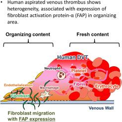
Fibroblast activation protein-α (FAP), a type-II transmembrane serine protease, is associated with wound healing, cancer-associated fibroblasts, and chronic fibrosing diseases. However, its expression in deep vein thrombosis (DVT) remains unclear. Therefore, this study investigated FAP expression and localization in DVT.
Methods
We performed pathological analyses of the aspirated thrombi of patients with DVT (n = 14), classifying thrombotic areas in terms of fresh, cellular lysis, and organizing reaction components. The organizing reaction included endothelialization and fibroblastic reaction. We immunohistochemically examined FAP-expressed areas and cells, and finally analyzed FAP expression in cultured dermal fibroblasts.
Results
All the aspirated thrombi showed a heterogeneous mixture of at least two of the three thrombotic areas. Specifically, 83 % of aspirated thrombi showed fresh and organizing reaction components. Immunohistochemical expression of FAP was restricted to the organizing area. Double immunofluorescence staining showed that FAP in the thrombi was mainly expressed in vimentin-positive or α-smooth muscle actin-positive fibroblasts. Some CD163-positive macrophages expressed FAP. FAP mRNA and protein levels were higher in fibroblasts with low-proliferative activity cultured under 0.1 % fetal bovine serum (FBS) than that under 10 % FBS. Fibroblasts cultured in 10 % FBS showed a significant decrease in FAP mRNA levels following supplementation with hemin, but not with thrombin.
Conclusions
The heterogeneous composition of venous thrombi suggests a multistep thrombus formation process in human DVT. Further, fibroblasts or myofibroblasts may express FAP during the organizing process. FAP expression may be higher in fibroblasts with low proliferative activity.
期刊介绍:
Thrombosis Research is an international journal dedicated to the swift dissemination of new information on thrombosis, hemostasis, and vascular biology, aimed at advancing both science and clinical care. The journal publishes peer-reviewed original research, reviews, editorials, opinions, and critiques, covering both basic and clinical studies. Priority is given to research that promises novel approaches in the diagnosis, therapy, prognosis, and prevention of thrombotic and hemorrhagic diseases.
 求助内容:
求助内容: 应助结果提醒方式:


