Natalie N. Khalil , Megan L. Rexius-Hall , Sean Escopete , Sarah J. Parker , Megan L. McCain
{"title":"急性缺氧和 TGF-β1 在人类成人心脏成纤维细胞中诱导的不同表型","authors":"Natalie N. Khalil , Megan L. Rexius-Hall , Sean Escopete , Sarah J. Parker , Megan L. McCain","doi":"10.1016/j.jmccpl.2024.100080","DOIUrl":null,"url":null,"abstract":"<div><p>Myocardial infarction (MI) causes hypoxic injury to downstream myocardial tissue, which initiates a wound healing response that replaces injured myocardial tissue with a scar. Wound healing is a complex process that consists of multiple phases, in which many different stimuli induce cardiac fibroblasts to differentiate into myofibroblasts and deposit new matrix. While this process is necessary to replace necrotic tissue, excessive and unresolved fibrosis is common post-MI and correlated with heart failure. Therefore, defining how cardiac fibroblast phenotypes are distinctly regulated by stimuli that are prevalent in the post-MI microenvironment, such as hypoxia and transforming growth factor-beta (TGF-β), is essential for understanding and ultimately mitigating pathological fibrosis. In this study, we acutely treated primary human adult cardiac fibroblasts with TGF-β1 or hypoxia and then characterized their phenotype through immunofluorescence, quantitative RT-PCR, and proteomic analysis. We found that fibroblasts responded to low oxygen with increased localization of hypoxia inducible factor 1 (HIF-1) to the nuclei after 4 h, which was followed by increased gene expression of vascular endothelial growth factor A (VEGFA), a known target of HIF-1, by 24 h. Both TGF-β1 and hypoxia inhibited proliferation after 24 h. TGF-β1 treatment also upregulated various fibrotic pathways. In contrast, hypoxia caused a reduction in several protein synthesis pathways, including collagen biosynthesis. Collectively, these data suggest that TGF-β1, but not acute hypoxia, robustly induces the differentiation of human cardiac fibroblasts into myofibroblasts. Discerning the overlapping and distinctive outcomes of TGF-β1 and hypoxia treatment is important for elucidating their roles in fibrotic remodeling post-MI and provides insight into potential therapeutic targets.</p></div>","PeriodicalId":73835,"journal":{"name":"Journal of molecular and cellular cardiology plus","volume":"9 ","pages":"Article 100080"},"PeriodicalIF":0.0000,"publicationDate":"2024-06-25","publicationTypes":"Journal Article","fieldsOfStudy":null,"isOpenAccess":false,"openAccessPdf":"https://www.sciencedirect.com/science/article/pii/S2772976124000205/pdfft?md5=aa347495a85d18d0d2f9e465fad8296c&pid=1-s2.0-S2772976124000205-main.pdf","citationCount":"0","resultStr":"{\"title\":\"Distinct phenotypes induced by acute hypoxia and TGF-β1 in human adult cardiac fibroblasts\",\"authors\":\"Natalie N. Khalil , Megan L. Rexius-Hall , Sean Escopete , Sarah J. Parker , Megan L. McCain\",\"doi\":\"10.1016/j.jmccpl.2024.100080\",\"DOIUrl\":null,\"url\":null,\"abstract\":\"<div><p>Myocardial infarction (MI) causes hypoxic injury to downstream myocardial tissue, which initiates a wound healing response that replaces injured myocardial tissue with a scar. Wound healing is a complex process that consists of multiple phases, in which many different stimuli induce cardiac fibroblasts to differentiate into myofibroblasts and deposit new matrix. While this process is necessary to replace necrotic tissue, excessive and unresolved fibrosis is common post-MI and correlated with heart failure. Therefore, defining how cardiac fibroblast phenotypes are distinctly regulated by stimuli that are prevalent in the post-MI microenvironment, such as hypoxia and transforming growth factor-beta (TGF-β), is essential for understanding and ultimately mitigating pathological fibrosis. In this study, we acutely treated primary human adult cardiac fibroblasts with TGF-β1 or hypoxia and then characterized their phenotype through immunofluorescence, quantitative RT-PCR, and proteomic analysis. We found that fibroblasts responded to low oxygen with increased localization of hypoxia inducible factor 1 (HIF-1) to the nuclei after 4 h, which was followed by increased gene expression of vascular endothelial growth factor A (VEGFA), a known target of HIF-1, by 24 h. Both TGF-β1 and hypoxia inhibited proliferation after 24 h. TGF-β1 treatment also upregulated various fibrotic pathways. In contrast, hypoxia caused a reduction in several protein synthesis pathways, including collagen biosynthesis. Collectively, these data suggest that TGF-β1, but not acute hypoxia, robustly induces the differentiation of human cardiac fibroblasts into myofibroblasts. Discerning the overlapping and distinctive outcomes of TGF-β1 and hypoxia treatment is important for elucidating their roles in fibrotic remodeling post-MI and provides insight into potential therapeutic targets.</p></div>\",\"PeriodicalId\":73835,\"journal\":{\"name\":\"Journal of molecular and cellular cardiology plus\",\"volume\":\"9 \",\"pages\":\"Article 100080\"},\"PeriodicalIF\":0.0000,\"publicationDate\":\"2024-06-25\",\"publicationTypes\":\"Journal Article\",\"fieldsOfStudy\":null,\"isOpenAccess\":false,\"openAccessPdf\":\"https://www.sciencedirect.com/science/article/pii/S2772976124000205/pdfft?md5=aa347495a85d18d0d2f9e465fad8296c&pid=1-s2.0-S2772976124000205-main.pdf\",\"citationCount\":\"0\",\"resultStr\":null,\"platform\":\"Semanticscholar\",\"paperid\":null,\"PeriodicalName\":\"Journal of molecular and cellular cardiology plus\",\"FirstCategoryId\":\"1085\",\"ListUrlMain\":\"https://www.sciencedirect.com/science/article/pii/S2772976124000205\",\"RegionNum\":0,\"RegionCategory\":null,\"ArticlePicture\":[],\"TitleCN\":null,\"AbstractTextCN\":null,\"PMCID\":null,\"EPubDate\":\"\",\"PubModel\":\"\",\"JCR\":\"\",\"JCRName\":\"\",\"Score\":null,\"Total\":0}","platform":"Semanticscholar","paperid":null,"PeriodicalName":"Journal of molecular and cellular cardiology plus","FirstCategoryId":"1085","ListUrlMain":"https://www.sciencedirect.com/science/article/pii/S2772976124000205","RegionNum":0,"RegionCategory":null,"ArticlePicture":[],"TitleCN":null,"AbstractTextCN":null,"PMCID":null,"EPubDate":"","PubModel":"","JCR":"","JCRName":"","Score":null,"Total":0}
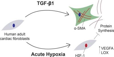
Distinct phenotypes induced by acute hypoxia and TGF-β1 in human adult cardiac fibroblasts
Myocardial infarction (MI) causes hypoxic injury to downstream myocardial tissue, which initiates a wound healing response that replaces injured myocardial tissue with a scar. Wound healing is a complex process that consists of multiple phases, in which many different stimuli induce cardiac fibroblasts to differentiate into myofibroblasts and deposit new matrix. While this process is necessary to replace necrotic tissue, excessive and unresolved fibrosis is common post-MI and correlated with heart failure. Therefore, defining how cardiac fibroblast phenotypes are distinctly regulated by stimuli that are prevalent in the post-MI microenvironment, such as hypoxia and transforming growth factor-beta (TGF-β), is essential for understanding and ultimately mitigating pathological fibrosis. In this study, we acutely treated primary human adult cardiac fibroblasts with TGF-β1 or hypoxia and then characterized their phenotype through immunofluorescence, quantitative RT-PCR, and proteomic analysis. We found that fibroblasts responded to low oxygen with increased localization of hypoxia inducible factor 1 (HIF-1) to the nuclei after 4 h, which was followed by increased gene expression of vascular endothelial growth factor A (VEGFA), a known target of HIF-1, by 24 h. Both TGF-β1 and hypoxia inhibited proliferation after 24 h. TGF-β1 treatment also upregulated various fibrotic pathways. In contrast, hypoxia caused a reduction in several protein synthesis pathways, including collagen biosynthesis. Collectively, these data suggest that TGF-β1, but not acute hypoxia, robustly induces the differentiation of human cardiac fibroblasts into myofibroblasts. Discerning the overlapping and distinctive outcomes of TGF-β1 and hypoxia treatment is important for elucidating their roles in fibrotic remodeling post-MI and provides insight into potential therapeutic targets.

 求助内容:
求助内容: 应助结果提醒方式:


