{"title":"Sp7 在成骨细胞增殖、分化和成骨过程形成中的作用","authors":"Qing Jiang , Kenichi Nagano , Takeshi Moriishi , Hisato Komori , Chiharu Sakane , Yuki Matsuo , Zhiguo Zhang , Riko Nishimura , Kosei Ito , Xin Qin , Toshihisa Komori","doi":"10.1016/j.jot.2024.06.005","DOIUrl":null,"url":null,"abstract":"<div><h3>Background</h3><p>Zinc finger-containing transcription factor Osterix/Specificity protein-7 (Sp7) is an essential transcription factor for osteoblast differentiation. However, its functions in differentiated osteoblasts remain unclear and the effects of osteoblast-specific <em>Sp7</em> deletion on osteocytes have not been sufficiently studied.</p></div><div><h3>Methods</h3><p><em>Sp7</em><sup>floxneo/floxneo</sup> mice, in which <em>Sp7</em> expression was 30 % of that in wild-type mice because of disturbed splicing by neo gene insertion, and osteoblast-specific knockout (<em>Sp7</em><sup>fl/fl;<em>Col1a1</em>−Cre</sup>) mice using 2.3-kb <em>Col1a1</em> enhanced green fluorescent protein (EGFP)-Cre were examined by micro-computed tomography (micro-CT), bone histomorphometry, serum markers, and histological analyses. The expression of osteoblast and osteocyte marker genes was examined by real-time reverse transcription (RT)-PCR analysis. Osteoblastogenesis, osteoclastogenesis, and regulation of the expression of collagen type I alpha 1 chain (<em>Col1a1</em>) were examined in primary osteoblasts.</p></div><div><h3>Results</h3><p>Femoral trabecular bone volume was higher in female <em>Sp7</em><sup>floxneo/floxneo</sup> and <em>Sp7</em><sup>fl/fl;<em>Col1a1</em>−Cre</sup> mice than in the respective controls, but not in males. Bromodeoxyuridine (BrdU)-positive osteoblastic cells were increased in male <em>Sp7</em><sup>fl/fl;<em>Col1a1</em>−Cre</sup> mice, and osteoblast number and the bone formation rate were increased in tibial trabecular bone in female <em>Sp7</em><sup>fl/fl;<em>Col1a1</em>−Cre</sup> mice, although osteoblast maturation was inhibited in female <em>Sp7</em><sup>fl/fl;<em>Col1a1</em>−Cre</sup> mice as shown by the increased expression of an immature osteoblast marker gene, secreted phosphoprotein 1 (<em>Spp1</em>), and reduced expression of a mature osteoblast marker gene, bone gamma-carboxyglutamate protein/bone gamma-carboxyglutamate protein 2 (<em>Bglap/Bglap2</em>). Furthermore, alkaline phosphatase activity was increased but mineralization was reduced in the culture of primary osteoblasts from <em>Sp7</em><sup>fl/fl;<em>Col1a1</em>−Cre</sup> mice. Therefore, the accumulated immature osteoblasts in <em>Sp7</em><sup>fl/fl;<em>Col1a1</em>−Cre</sup> mice was likely compensated for the inhibition of osteoblast maturation at different levels in males and females. Vertebral trabecular bone volume was lower in both male and female <em>Sp7</em><sup>fl/fl;<em>Col1a1</em>−Cre</sup> mice than in the controls and the osteoblast parameters and bone formation rate in females were lower in <em>Sp7</em><sup>fl/fl;<em>Col1a1</em>−Cre</sup> mice than in <em>Sp7</em><sup>fl/fl</sup> mice, suggesting differential regulatory mechanisms in long bones and vertebrae. The femoral cortical bone was thin and porous in <em>Sp7</em><sup>floxneo/floxneo</sup> and <em>Sp7</em><sup>fl/fl;<em>Col1a1</em>−Cre</sup> mice of both sexes, the number of canaliculi was reduced, and terminal deoxynucleotidyl transferase-mediated dUTP nick end labelling (TUNEL)-positive lacunae and the osteoclasts were increased, whereas the bone formation rate was similar in <em>Sp7</em><sup>fl/fl;<em>Col1a1</em>−Cre</sup> and <em>Sp7</em><sup>fl/fl</sup> mice. The serum levels of total procollagen type 1 N-terminal propeptide (P1NP), a marker for bone formation, were similar, while those of tartrate-resistant acid phosphatase 5b (TRAP5b), a marker for bone resorption, were higher in <em>Sp7</em><sup>fl/fl;<em>Col1a1</em>−Cre</sup> mice. Osteoblasts were less cuboidal, the expression of <em>Col1a1</em> and <em>Col1a1</em>-EGFP-Cre was lower in <em>Sp7</em><sup>fl/fl;<em>Col1a1</em>−Cre</sup> mice, and overexpression of <em>Sp7</em> induced <em>Col1a1</em> expression.</p></div><div><h3>Conclusions</h3><p>Our studies indicated that Sp7 inhibits the proliferation of immature osteoblasts, induces osteoblast maturation and <em>Col1a1</em> expression, and is required for osteocytes to acquire a sufficient number of processes for their survival, which prevents cortical porosity.</p></div><div><h3>The translational potential of this article</h3><p>This study clarified the roles of Sp7 in differentiated osteoblasts in proliferarion, maturation, <em>Col1a1</em> expression, and osteocyte process formation, which are required for targeting SP7 in the development of therapies for osteoporosis.</p></div>","PeriodicalId":16636,"journal":{"name":"Journal of Orthopaedic Translation","volume":"47 ","pages":"Pages 161-175"},"PeriodicalIF":5.9000,"publicationDate":"2024-07-01","publicationTypes":"Journal Article","fieldsOfStudy":null,"isOpenAccess":false,"openAccessPdf":"https://www.sciencedirect.com/science/article/pii/S2214031X24000536/pdfft?md5=fcf3b3db8cc860edc417dcaa8f83f032&pid=1-s2.0-S2214031X24000536-main.pdf","citationCount":"0","resultStr":"{\"title\":\"Roles of Sp7 in osteoblasts for the proliferation, differentiation, and osteocyte process formation\",\"authors\":\"Qing Jiang , Kenichi Nagano , Takeshi Moriishi , Hisato Komori , Chiharu Sakane , Yuki Matsuo , Zhiguo Zhang , Riko Nishimura , Kosei Ito , Xin Qin , Toshihisa Komori\",\"doi\":\"10.1016/j.jot.2024.06.005\",\"DOIUrl\":null,\"url\":null,\"abstract\":\"<div><h3>Background</h3><p>Zinc finger-containing transcription factor Osterix/Specificity protein-7 (Sp7) is an essential transcription factor for osteoblast differentiation. However, its functions in differentiated osteoblasts remain unclear and the effects of osteoblast-specific <em>Sp7</em> deletion on osteocytes have not been sufficiently studied.</p></div><div><h3>Methods</h3><p><em>Sp7</em><sup>floxneo/floxneo</sup> mice, in which <em>Sp7</em> expression was 30 % of that in wild-type mice because of disturbed splicing by neo gene insertion, and osteoblast-specific knockout (<em>Sp7</em><sup>fl/fl;<em>Col1a1</em>−Cre</sup>) mice using 2.3-kb <em>Col1a1</em> enhanced green fluorescent protein (EGFP)-Cre were examined by micro-computed tomography (micro-CT), bone histomorphometry, serum markers, and histological analyses. The expression of osteoblast and osteocyte marker genes was examined by real-time reverse transcription (RT)-PCR analysis. Osteoblastogenesis, osteoclastogenesis, and regulation of the expression of collagen type I alpha 1 chain (<em>Col1a1</em>) were examined in primary osteoblasts.</p></div><div><h3>Results</h3><p>Femoral trabecular bone volume was higher in female <em>Sp7</em><sup>floxneo/floxneo</sup> and <em>Sp7</em><sup>fl/fl;<em>Col1a1</em>−Cre</sup> mice than in the respective controls, but not in males. Bromodeoxyuridine (BrdU)-positive osteoblastic cells were increased in male <em>Sp7</em><sup>fl/fl;<em>Col1a1</em>−Cre</sup> mice, and osteoblast number and the bone formation rate were increased in tibial trabecular bone in female <em>Sp7</em><sup>fl/fl;<em>Col1a1</em>−Cre</sup> mice, although osteoblast maturation was inhibited in female <em>Sp7</em><sup>fl/fl;<em>Col1a1</em>−Cre</sup> mice as shown by the increased expression of an immature osteoblast marker gene, secreted phosphoprotein 1 (<em>Spp1</em>), and reduced expression of a mature osteoblast marker gene, bone gamma-carboxyglutamate protein/bone gamma-carboxyglutamate protein 2 (<em>Bglap/Bglap2</em>). Furthermore, alkaline phosphatase activity was increased but mineralization was reduced in the culture of primary osteoblasts from <em>Sp7</em><sup>fl/fl;<em>Col1a1</em>−Cre</sup> mice. Therefore, the accumulated immature osteoblasts in <em>Sp7</em><sup>fl/fl;<em>Col1a1</em>−Cre</sup> mice was likely compensated for the inhibition of osteoblast maturation at different levels in males and females. Vertebral trabecular bone volume was lower in both male and female <em>Sp7</em><sup>fl/fl;<em>Col1a1</em>−Cre</sup> mice than in the controls and the osteoblast parameters and bone formation rate in females were lower in <em>Sp7</em><sup>fl/fl;<em>Col1a1</em>−Cre</sup> mice than in <em>Sp7</em><sup>fl/fl</sup> mice, suggesting differential regulatory mechanisms in long bones and vertebrae. The femoral cortical bone was thin and porous in <em>Sp7</em><sup>floxneo/floxneo</sup> and <em>Sp7</em><sup>fl/fl;<em>Col1a1</em>−Cre</sup> mice of both sexes, the number of canaliculi was reduced, and terminal deoxynucleotidyl transferase-mediated dUTP nick end labelling (TUNEL)-positive lacunae and the osteoclasts were increased, whereas the bone formation rate was similar in <em>Sp7</em><sup>fl/fl;<em>Col1a1</em>−Cre</sup> and <em>Sp7</em><sup>fl/fl</sup> mice. The serum levels of total procollagen type 1 N-terminal propeptide (P1NP), a marker for bone formation, were similar, while those of tartrate-resistant acid phosphatase 5b (TRAP5b), a marker for bone resorption, were higher in <em>Sp7</em><sup>fl/fl;<em>Col1a1</em>−Cre</sup> mice. Osteoblasts were less cuboidal, the expression of <em>Col1a1</em> and <em>Col1a1</em>-EGFP-Cre was lower in <em>Sp7</em><sup>fl/fl;<em>Col1a1</em>−Cre</sup> mice, and overexpression of <em>Sp7</em> induced <em>Col1a1</em> expression.</p></div><div><h3>Conclusions</h3><p>Our studies indicated that Sp7 inhibits the proliferation of immature osteoblasts, induces osteoblast maturation and <em>Col1a1</em> expression, and is required for osteocytes to acquire a sufficient number of processes for their survival, which prevents cortical porosity.</p></div><div><h3>The translational potential of this article</h3><p>This study clarified the roles of Sp7 in differentiated osteoblasts in proliferarion, maturation, <em>Col1a1</em> expression, and osteocyte process formation, which are required for targeting SP7 in the development of therapies for osteoporosis.</p></div>\",\"PeriodicalId\":16636,\"journal\":{\"name\":\"Journal of Orthopaedic Translation\",\"volume\":\"47 \",\"pages\":\"Pages 161-175\"},\"PeriodicalIF\":5.9000,\"publicationDate\":\"2024-07-01\",\"publicationTypes\":\"Journal Article\",\"fieldsOfStudy\":null,\"isOpenAccess\":false,\"openAccessPdf\":\"https://www.sciencedirect.com/science/article/pii/S2214031X24000536/pdfft?md5=fcf3b3db8cc860edc417dcaa8f83f032&pid=1-s2.0-S2214031X24000536-main.pdf\",\"citationCount\":\"0\",\"resultStr\":null,\"platform\":\"Semanticscholar\",\"paperid\":null,\"PeriodicalName\":\"Journal of Orthopaedic Translation\",\"FirstCategoryId\":\"3\",\"ListUrlMain\":\"https://www.sciencedirect.com/science/article/pii/S2214031X24000536\",\"RegionNum\":1,\"RegionCategory\":\"医学\",\"ArticlePicture\":[],\"TitleCN\":null,\"AbstractTextCN\":null,\"PMCID\":null,\"EPubDate\":\"\",\"PubModel\":\"\",\"JCR\":\"Q1\",\"JCRName\":\"ORTHOPEDICS\",\"Score\":null,\"Total\":0}","platform":"Semanticscholar","paperid":null,"PeriodicalName":"Journal of Orthopaedic Translation","FirstCategoryId":"3","ListUrlMain":"https://www.sciencedirect.com/science/article/pii/S2214031X24000536","RegionNum":1,"RegionCategory":"医学","ArticlePicture":[],"TitleCN":null,"AbstractTextCN":null,"PMCID":null,"EPubDate":"","PubModel":"","JCR":"Q1","JCRName":"ORTHOPEDICS","Score":null,"Total":0}
Roles of Sp7 in osteoblasts for the proliferation, differentiation, and osteocyte process formation
Background
Zinc finger-containing transcription factor Osterix/Specificity protein-7 (Sp7) is an essential transcription factor for osteoblast differentiation. However, its functions in differentiated osteoblasts remain unclear and the effects of osteoblast-specific Sp7 deletion on osteocytes have not been sufficiently studied.
Methods
Sp7floxneo/floxneo mice, in which Sp7 expression was 30 % of that in wild-type mice because of disturbed splicing by neo gene insertion, and osteoblast-specific knockout (Sp7fl/fl;Col1a1−Cre) mice using 2.3-kb Col1a1 enhanced green fluorescent protein (EGFP)-Cre were examined by micro-computed tomography (micro-CT), bone histomorphometry, serum markers, and histological analyses. The expression of osteoblast and osteocyte marker genes was examined by real-time reverse transcription (RT)-PCR analysis. Osteoblastogenesis, osteoclastogenesis, and regulation of the expression of collagen type I alpha 1 chain (Col1a1) were examined in primary osteoblasts.
Results
Femoral trabecular bone volume was higher in female Sp7floxneo/floxneo and Sp7fl/fl;Col1a1−Cre mice than in the respective controls, but not in males. Bromodeoxyuridine (BrdU)-positive osteoblastic cells were increased in male Sp7fl/fl;Col1a1−Cre mice, and osteoblast number and the bone formation rate were increased in tibial trabecular bone in female Sp7fl/fl;Col1a1−Cre mice, although osteoblast maturation was inhibited in female Sp7fl/fl;Col1a1−Cre mice as shown by the increased expression of an immature osteoblast marker gene, secreted phosphoprotein 1 (Spp1), and reduced expression of a mature osteoblast marker gene, bone gamma-carboxyglutamate protein/bone gamma-carboxyglutamate protein 2 (Bglap/Bglap2). Furthermore, alkaline phosphatase activity was increased but mineralization was reduced in the culture of primary osteoblasts from Sp7fl/fl;Col1a1−Cre mice. Therefore, the accumulated immature osteoblasts in Sp7fl/fl;Col1a1−Cre mice was likely compensated for the inhibition of osteoblast maturation at different levels in males and females. Vertebral trabecular bone volume was lower in both male and female Sp7fl/fl;Col1a1−Cre mice than in the controls and the osteoblast parameters and bone formation rate in females were lower in Sp7fl/fl;Col1a1−Cre mice than in Sp7fl/fl mice, suggesting differential regulatory mechanisms in long bones and vertebrae. The femoral cortical bone was thin and porous in Sp7floxneo/floxneo and Sp7fl/fl;Col1a1−Cre mice of both sexes, the number of canaliculi was reduced, and terminal deoxynucleotidyl transferase-mediated dUTP nick end labelling (TUNEL)-positive lacunae and the osteoclasts were increased, whereas the bone formation rate was similar in Sp7fl/fl;Col1a1−Cre and Sp7fl/fl mice. The serum levels of total procollagen type 1 N-terminal propeptide (P1NP), a marker for bone formation, were similar, while those of tartrate-resistant acid phosphatase 5b (TRAP5b), a marker for bone resorption, were higher in Sp7fl/fl;Col1a1−Cre mice. Osteoblasts were less cuboidal, the expression of Col1a1 and Col1a1-EGFP-Cre was lower in Sp7fl/fl;Col1a1−Cre mice, and overexpression of Sp7 induced Col1a1 expression.
Conclusions
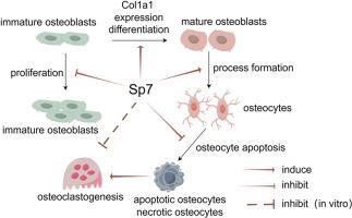
Our studies indicated that Sp7 inhibits the proliferation of immature osteoblasts, induces osteoblast maturation and Col1a1 expression, and is required for osteocytes to acquire a sufficient number of processes for their survival, which prevents cortical porosity.
The translational potential of this article
This study clarified the roles of Sp7 in differentiated osteoblasts in proliferarion, maturation, Col1a1 expression, and osteocyte process formation, which are required for targeting SP7 in the development of therapies for osteoporosis.
期刊介绍:
The Journal of Orthopaedic Translation (JOT) is the official peer-reviewed, open access journal of the Chinese Speaking Orthopaedic Society (CSOS) and the International Chinese Musculoskeletal Research Society (ICMRS). It is published quarterly, in January, April, July and October, by Elsevier.
 求助内容:
求助内容: 应助结果提醒方式:


