{"title":"对比增强 FLAIR 成像在诊断小儿葡萄膜炎中的实用性。","authors":"Saffet Ozturk, Esin Kurtulus Ozturk, Yasemin Tasci Yildiz, Bahadir Konuskan","doi":"10.5334/jbsr.3565","DOIUrl":null,"url":null,"abstract":"<p><p><i>Objectives:</i> Contrast-enhanced FLAIR fat-suppressed (CE-FLAIR-FS) imaging can potentially increase the diagnostic accuracy of uveal diseases and ultimately provide better patient management. This study aimed to determine the diagnostic value of CE-FLAIR-FS imaging versus contrast-enhanced T1-weighted imaging (CE-T1WI) in the assessment of pediatric patients with uveitis. <i>Material and methods:</i> Twenty-one children with uveitis who underwent whole brain magnetic resonance imaging (MRI), including CE-FLAIR-FS and CE-T1WI, were retrospectively included in the study. We evaluated the presence of uveal tract contrast enhancement with thickening, vitreous humor signal abnormality, and accompanying brain abnormalities. The uveal enhancement intensity was assessed semiquantitatively as mild, moderate, and marked uveitis compared to CE-T1WI and CE-FLAIR-FS images. <i>Results:</i> Panuveitis (61.9%) was the most frequent anatomic location, and most of them were idiopathic (47.6%). Of the 42 eyes with clinical uveitis, enhancement of the uveal tract was observed on CE-FLAIR-FS images in 21 eyes (50%), while in 5 eyes (11.9%) on CE-T1WI. The sensitivity of CE-FLAIR-FS in panuveitis was detected to be quite high (80.8%). The number of affected eyes and enhancement degree were found to be higher on CE-FLAIR-FS (<i>p</i> < 0.001). In assessing the severity of uveitis, CE-FLAIR-FS grades were significantly higher and more sensitive than CE-T1WI (<i>p</i> < 0.001, <i>Z</i>: -4.347). Three patients had vitreous abnormal signals on CE-FLAIR-FS images, but none on CE-T1WI. <i>Conclusion:</i> CE-FLAIR-FS plays a significant role in the diagnosis of pediatric uveitis, identifying the involvement and severity of the uveal inflammation and guiding the appropriate management. It would be beneficial to add it as a standard sequence to the routine MRI protocol for uveal pathologies.</p>","PeriodicalId":55987,"journal":{"name":"Journal of the Belgian Society of Radiology","volume":"108 1","pages":"66"},"PeriodicalIF":1.3000,"publicationDate":"2024-06-28","publicationTypes":"Journal Article","fieldsOfStudy":null,"isOpenAccess":false,"openAccessPdf":"https://www.ncbi.nlm.nih.gov/pmc/articles/PMC11212780/pdf/","citationCount":"0","resultStr":"{\"title\":\"The Diagnostic Utility of Contrast-Enhanced FLAIR Imaging in the Diagnosis of Pediatric Uveitis.\",\"authors\":\"Saffet Ozturk, Esin Kurtulus Ozturk, Yasemin Tasci Yildiz, Bahadir Konuskan\",\"doi\":\"10.5334/jbsr.3565\",\"DOIUrl\":null,\"url\":null,\"abstract\":\"<p><p><i>Objectives:</i> Contrast-enhanced FLAIR fat-suppressed (CE-FLAIR-FS) imaging can potentially increase the diagnostic accuracy of uveal diseases and ultimately provide better patient management. This study aimed to determine the diagnostic value of CE-FLAIR-FS imaging versus contrast-enhanced T1-weighted imaging (CE-T1WI) in the assessment of pediatric patients with uveitis. <i>Material and methods:</i> Twenty-one children with uveitis who underwent whole brain magnetic resonance imaging (MRI), including CE-FLAIR-FS and CE-T1WI, were retrospectively included in the study. We evaluated the presence of uveal tract contrast enhancement with thickening, vitreous humor signal abnormality, and accompanying brain abnormalities. The uveal enhancement intensity was assessed semiquantitatively as mild, moderate, and marked uveitis compared to CE-T1WI and CE-FLAIR-FS images. <i>Results:</i> Panuveitis (61.9%) was the most frequent anatomic location, and most of them were idiopathic (47.6%). Of the 42 eyes with clinical uveitis, enhancement of the uveal tract was observed on CE-FLAIR-FS images in 21 eyes (50%), while in 5 eyes (11.9%) on CE-T1WI. The sensitivity of CE-FLAIR-FS in panuveitis was detected to be quite high (80.8%). The number of affected eyes and enhancement degree were found to be higher on CE-FLAIR-FS (<i>p</i> < 0.001). In assessing the severity of uveitis, CE-FLAIR-FS grades were significantly higher and more sensitive than CE-T1WI (<i>p</i> < 0.001, <i>Z</i>: -4.347). Three patients had vitreous abnormal signals on CE-FLAIR-FS images, but none on CE-T1WI. <i>Conclusion:</i> CE-FLAIR-FS plays a significant role in the diagnosis of pediatric uveitis, identifying the involvement and severity of the uveal inflammation and guiding the appropriate management. It would be beneficial to add it as a standard sequence to the routine MRI protocol for uveal pathologies.</p>\",\"PeriodicalId\":55987,\"journal\":{\"name\":\"Journal of the Belgian Society of Radiology\",\"volume\":\"108 1\",\"pages\":\"66\"},\"PeriodicalIF\":1.3000,\"publicationDate\":\"2024-06-28\",\"publicationTypes\":\"Journal Article\",\"fieldsOfStudy\":null,\"isOpenAccess\":false,\"openAccessPdf\":\"https://www.ncbi.nlm.nih.gov/pmc/articles/PMC11212780/pdf/\",\"citationCount\":\"0\",\"resultStr\":null,\"platform\":\"Semanticscholar\",\"paperid\":null,\"PeriodicalName\":\"Journal of the Belgian Society of Radiology\",\"FirstCategoryId\":\"3\",\"ListUrlMain\":\"https://doi.org/10.5334/jbsr.3565\",\"RegionNum\":4,\"RegionCategory\":\"医学\",\"ArticlePicture\":[],\"TitleCN\":null,\"AbstractTextCN\":null,\"PMCID\":null,\"EPubDate\":\"2024/1/1 0:00:00\",\"PubModel\":\"eCollection\",\"JCR\":\"Q4\",\"JCRName\":\"RADIOLOGY, NUCLEAR MEDICINE & MEDICAL IMAGING\",\"Score\":null,\"Total\":0}","platform":"Semanticscholar","paperid":null,"PeriodicalName":"Journal of the Belgian Society of Radiology","FirstCategoryId":"3","ListUrlMain":"https://doi.org/10.5334/jbsr.3565","RegionNum":4,"RegionCategory":"医学","ArticlePicture":[],"TitleCN":null,"AbstractTextCN":null,"PMCID":null,"EPubDate":"2024/1/1 0:00:00","PubModel":"eCollection","JCR":"Q4","JCRName":"RADIOLOGY, NUCLEAR MEDICINE & MEDICAL IMAGING","Score":null,"Total":0}
The Diagnostic Utility of Contrast-Enhanced FLAIR Imaging in the Diagnosis of Pediatric Uveitis.
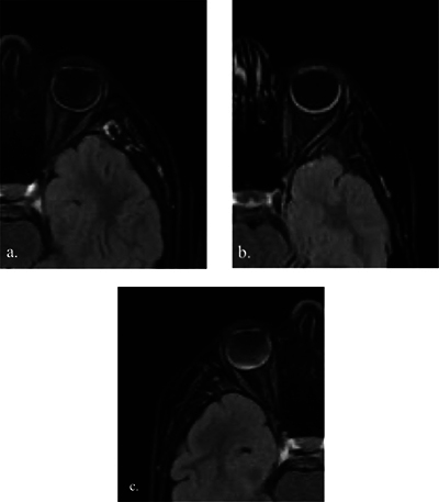
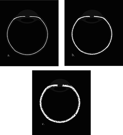
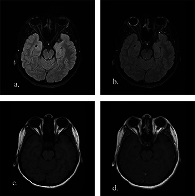
Objectives: Contrast-enhanced FLAIR fat-suppressed (CE-FLAIR-FS) imaging can potentially increase the diagnostic accuracy of uveal diseases and ultimately provide better patient management. This study aimed to determine the diagnostic value of CE-FLAIR-FS imaging versus contrast-enhanced T1-weighted imaging (CE-T1WI) in the assessment of pediatric patients with uveitis. Material and methods: Twenty-one children with uveitis who underwent whole brain magnetic resonance imaging (MRI), including CE-FLAIR-FS and CE-T1WI, were retrospectively included in the study. We evaluated the presence of uveal tract contrast enhancement with thickening, vitreous humor signal abnormality, and accompanying brain abnormalities. The uveal enhancement intensity was assessed semiquantitatively as mild, moderate, and marked uveitis compared to CE-T1WI and CE-FLAIR-FS images. Results: Panuveitis (61.9%) was the most frequent anatomic location, and most of them were idiopathic (47.6%). Of the 42 eyes with clinical uveitis, enhancement of the uveal tract was observed on CE-FLAIR-FS images in 21 eyes (50%), while in 5 eyes (11.9%) on CE-T1WI. The sensitivity of CE-FLAIR-FS in panuveitis was detected to be quite high (80.8%). The number of affected eyes and enhancement degree were found to be higher on CE-FLAIR-FS (p < 0.001). In assessing the severity of uveitis, CE-FLAIR-FS grades were significantly higher and more sensitive than CE-T1WI (p < 0.001, Z: -4.347). Three patients had vitreous abnormal signals on CE-FLAIR-FS images, but none on CE-T1WI. Conclusion: CE-FLAIR-FS plays a significant role in the diagnosis of pediatric uveitis, identifying the involvement and severity of the uveal inflammation and guiding the appropriate management. It would be beneficial to add it as a standard sequence to the routine MRI protocol for uveal pathologies.
期刊介绍:
The purpose of the Journal of the Belgian Society of Radiology is the publication of articles dealing with diagnostic and interventional radiology, related imaging techniques, allied sciences, and continuing education.
 求助内容:
求助内容: 应助结果提醒方式:


