Xuanxuan Zhang , Jicong Chen , Ruohui Lin , Yaping Huang , Ziyuan Wang , Susu Xu , Lei Wang , Fang Chen , Jian Zhang , Ke Pan , Zhiqi Yin
{"title":"乳酸盐通过 H3K14la/KLF5 通路驱动糖尿病肾病的上皮-间质转化","authors":"Xuanxuan Zhang , Jicong Chen , Ruohui Lin , Yaping Huang , Ziyuan Wang , Susu Xu , Lei Wang , Fang Chen , Jian Zhang , Ke Pan , Zhiqi Yin","doi":"10.1016/j.redox.2024.103246","DOIUrl":null,"url":null,"abstract":"<div><p>High levels of urinary lactate are an increased risk of progression in patients with diabetic kidney disease (DKD). However, it is still unveiled how lactate drive DKD. Epithelial-mesenchymal transition (EMT), which is characterized by the loss of epithelial cells polarity and cell-cell adhesion, and the acquisition of mesenchymal-like phenotypes, is widely recognized a critical contributor to DKD. Here, we found a switch from oxidative phosphorylation (OXPHOS) toward glycolysis in AGEs-induced renal tubular epithelial cells, thus leading to elevated levels of renal lactic acid. We demonstrated that reducing the lactate levels markedly delayed EMT progression and improved renal tubular fibrosis in DKD. Mechanically, we observed lactate increased the levels of histone H3 lysine 14 lactylation (H3K14la) in DKD. ChIP-seq & RNA-seq results showed histone lactylation contributed to EMT process by facilitating KLF5 expression. Moreover, KLF5 recognized the promotor of cdh1 and inhibited its transcription, which accelerated EMT of DKD. Additionally, nephro-specific knockdown and pharmacological inhibition of KLF5 diminished EMT development and attenuated DKD fibrosis. Thus, our study provides better understanding of epigenetic regulation of DKD pathogenesis, and new therapeutic strategy for DKD by disruption of the lactate-drived H3K14la/KLF5 pathway.</p></div>","PeriodicalId":20998,"journal":{"name":"Redox Biology","volume":null,"pages":null},"PeriodicalIF":10.7000,"publicationDate":"2024-06-20","publicationTypes":"Journal Article","fieldsOfStudy":null,"isOpenAccess":false,"openAccessPdf":"https://www.sciencedirect.com/science/article/pii/S2213231724002246/pdfft?md5=2328d84588f52c9562c16deba5fec867&pid=1-s2.0-S2213231724002246-main.pdf","citationCount":"0","resultStr":"{\"title\":\"Lactate drives epithelial-mesenchymal transition in diabetic kidney disease via the H3K14la/KLF5 pathway\",\"authors\":\"Xuanxuan Zhang , Jicong Chen , Ruohui Lin , Yaping Huang , Ziyuan Wang , Susu Xu , Lei Wang , Fang Chen , Jian Zhang , Ke Pan , Zhiqi Yin\",\"doi\":\"10.1016/j.redox.2024.103246\",\"DOIUrl\":null,\"url\":null,\"abstract\":\"<div><p>High levels of urinary lactate are an increased risk of progression in patients with diabetic kidney disease (DKD). However, it is still unveiled how lactate drive DKD. Epithelial-mesenchymal transition (EMT), which is characterized by the loss of epithelial cells polarity and cell-cell adhesion, and the acquisition of mesenchymal-like phenotypes, is widely recognized a critical contributor to DKD. Here, we found a switch from oxidative phosphorylation (OXPHOS) toward glycolysis in AGEs-induced renal tubular epithelial cells, thus leading to elevated levels of renal lactic acid. We demonstrated that reducing the lactate levels markedly delayed EMT progression and improved renal tubular fibrosis in DKD. Mechanically, we observed lactate increased the levels of histone H3 lysine 14 lactylation (H3K14la) in DKD. ChIP-seq & RNA-seq results showed histone lactylation contributed to EMT process by facilitating KLF5 expression. Moreover, KLF5 recognized the promotor of cdh1 and inhibited its transcription, which accelerated EMT of DKD. Additionally, nephro-specific knockdown and pharmacological inhibition of KLF5 diminished EMT development and attenuated DKD fibrosis. Thus, our study provides better understanding of epigenetic regulation of DKD pathogenesis, and new therapeutic strategy for DKD by disruption of the lactate-drived H3K14la/KLF5 pathway.</p></div>\",\"PeriodicalId\":20998,\"journal\":{\"name\":\"Redox Biology\",\"volume\":null,\"pages\":null},\"PeriodicalIF\":10.7000,\"publicationDate\":\"2024-06-20\",\"publicationTypes\":\"Journal Article\",\"fieldsOfStudy\":null,\"isOpenAccess\":false,\"openAccessPdf\":\"https://www.sciencedirect.com/science/article/pii/S2213231724002246/pdfft?md5=2328d84588f52c9562c16deba5fec867&pid=1-s2.0-S2213231724002246-main.pdf\",\"citationCount\":\"0\",\"resultStr\":null,\"platform\":\"Semanticscholar\",\"paperid\":null,\"PeriodicalName\":\"Redox Biology\",\"FirstCategoryId\":\"99\",\"ListUrlMain\":\"https://www.sciencedirect.com/science/article/pii/S2213231724002246\",\"RegionNum\":1,\"RegionCategory\":\"生物学\",\"ArticlePicture\":[],\"TitleCN\":null,\"AbstractTextCN\":null,\"PMCID\":null,\"EPubDate\":\"\",\"PubModel\":\"\",\"JCR\":\"Q1\",\"JCRName\":\"BIOCHEMISTRY & MOLECULAR BIOLOGY\",\"Score\":null,\"Total\":0}","platform":"Semanticscholar","paperid":null,"PeriodicalName":"Redox Biology","FirstCategoryId":"99","ListUrlMain":"https://www.sciencedirect.com/science/article/pii/S2213231724002246","RegionNum":1,"RegionCategory":"生物学","ArticlePicture":[],"TitleCN":null,"AbstractTextCN":null,"PMCID":null,"EPubDate":"","PubModel":"","JCR":"Q1","JCRName":"BIOCHEMISTRY & MOLECULAR BIOLOGY","Score":null,"Total":0}
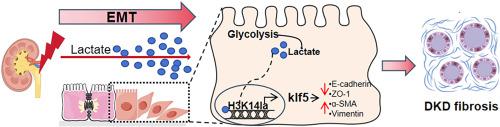
Lactate drives epithelial-mesenchymal transition in diabetic kidney disease via the H3K14la/KLF5 pathway
High levels of urinary lactate are an increased risk of progression in patients with diabetic kidney disease (DKD). However, it is still unveiled how lactate drive DKD. Epithelial-mesenchymal transition (EMT), which is characterized by the loss of epithelial cells polarity and cell-cell adhesion, and the acquisition of mesenchymal-like phenotypes, is widely recognized a critical contributor to DKD. Here, we found a switch from oxidative phosphorylation (OXPHOS) toward glycolysis in AGEs-induced renal tubular epithelial cells, thus leading to elevated levels of renal lactic acid. We demonstrated that reducing the lactate levels markedly delayed EMT progression and improved renal tubular fibrosis in DKD. Mechanically, we observed lactate increased the levels of histone H3 lysine 14 lactylation (H3K14la) in DKD. ChIP-seq & RNA-seq results showed histone lactylation contributed to EMT process by facilitating KLF5 expression. Moreover, KLF5 recognized the promotor of cdh1 and inhibited its transcription, which accelerated EMT of DKD. Additionally, nephro-specific knockdown and pharmacological inhibition of KLF5 diminished EMT development and attenuated DKD fibrosis. Thus, our study provides better understanding of epigenetic regulation of DKD pathogenesis, and new therapeutic strategy for DKD by disruption of the lactate-drived H3K14la/KLF5 pathway.
期刊介绍:
Redox Biology is the official journal of the Society for Redox Biology and Medicine and the Society for Free Radical Research-Europe. It is also affiliated with the International Society for Free Radical Research (SFRRI). This journal serves as a platform for publishing pioneering research, innovative methods, and comprehensive review articles in the field of redox biology, encompassing both health and disease.
Redox Biology welcomes various forms of contributions, including research articles (short or full communications), methods, mini-reviews, and commentaries. Through its diverse range of published content, Redox Biology aims to foster advancements and insights in the understanding of redox biology and its implications.

 求助内容:
求助内容: 应助结果提醒方式:


