M. A. Model, R. Guo, K. Fasina, R. Jin, R. J. Clements, L. G. Leff
{"title":"在透光显微镜下测量细菌和小型细胞器中的蛋白质浓度。","authors":"M. A. Model, R. Guo, K. Fasina, R. Jin, R. J. Clements, L. G. Leff","doi":"10.1002/jmr.3099","DOIUrl":null,"url":null,"abstract":"<p>Protein concentration (PC) is an essential characteristic of cells and organelles; it determines the extent of macromolecular crowding effects and serves as a sensitive indicator of cellular health. A simple and direct way to quantify PC is provided by brightfield-based transport-of-intensity equation (TIE) imaging combined with volume measurements. However, since TIE is based on geometric optics, its applicability to micrometer-sized particles is not clear. Here, we show that TIE can be used on particles with sizes comparable to the wavelength. At the same time, we introduce a new ImageJ plugin that allows TIE image processing without resorting to advanced mathematical programs. To convert TIE data to PC, knowledge of particle volumes is essential. The volumes of bacteria or other isolated particles can be measured by displacement of an external absorbing dye (“transmission-through-dye” or TTD microscopy), and for spherical intracellular particles, volumes can be estimated from their diameters. We illustrate the use of TIE on <i>Escherichia coli</i>, mammalian nucleoli, and nucleolar fibrillar centers. The method is easy to use and achieves high spatial resolution.</p>","PeriodicalId":16531,"journal":{"name":"Journal of Molecular Recognition","volume":"37 5","pages":""},"PeriodicalIF":3.0000,"publicationDate":"2024-06-25","publicationTypes":"Journal Article","fieldsOfStudy":null,"isOpenAccess":false,"openAccessPdf":"https://onlinelibrary.wiley.com/doi/epdf/10.1002/jmr.3099","citationCount":"0","resultStr":"{\"title\":\"Measurement of protein concentration in bacteria and small organelles under a light transmission microscope\",\"authors\":\"M. A. Model, R. Guo, K. Fasina, R. Jin, R. J. Clements, L. G. Leff\",\"doi\":\"10.1002/jmr.3099\",\"DOIUrl\":null,\"url\":null,\"abstract\":\"<p>Protein concentration (PC) is an essential characteristic of cells and organelles; it determines the extent of macromolecular crowding effects and serves as a sensitive indicator of cellular health. A simple and direct way to quantify PC is provided by brightfield-based transport-of-intensity equation (TIE) imaging combined with volume measurements. However, since TIE is based on geometric optics, its applicability to micrometer-sized particles is not clear. Here, we show that TIE can be used on particles with sizes comparable to the wavelength. At the same time, we introduce a new ImageJ plugin that allows TIE image processing without resorting to advanced mathematical programs. To convert TIE data to PC, knowledge of particle volumes is essential. The volumes of bacteria or other isolated particles can be measured by displacement of an external absorbing dye (“transmission-through-dye” or TTD microscopy), and for spherical intracellular particles, volumes can be estimated from their diameters. We illustrate the use of TIE on <i>Escherichia coli</i>, mammalian nucleoli, and nucleolar fibrillar centers. The method is easy to use and achieves high spatial resolution.</p>\",\"PeriodicalId\":16531,\"journal\":{\"name\":\"Journal of Molecular Recognition\",\"volume\":\"37 5\",\"pages\":\"\"},\"PeriodicalIF\":3.0000,\"publicationDate\":\"2024-06-25\",\"publicationTypes\":\"Journal Article\",\"fieldsOfStudy\":null,\"isOpenAccess\":false,\"openAccessPdf\":\"https://onlinelibrary.wiley.com/doi/epdf/10.1002/jmr.3099\",\"citationCount\":\"0\",\"resultStr\":null,\"platform\":\"Semanticscholar\",\"paperid\":null,\"PeriodicalName\":\"Journal of Molecular Recognition\",\"FirstCategoryId\":\"99\",\"ListUrlMain\":\"https://onlinelibrary.wiley.com/doi/10.1002/jmr.3099\",\"RegionNum\":4,\"RegionCategory\":\"生物学\",\"ArticlePicture\":[],\"TitleCN\":null,\"AbstractTextCN\":null,\"PMCID\":null,\"EPubDate\":\"\",\"PubModel\":\"\",\"JCR\":\"Q3\",\"JCRName\":\"BIOCHEMISTRY & MOLECULAR BIOLOGY\",\"Score\":null,\"Total\":0}","platform":"Semanticscholar","paperid":null,"PeriodicalName":"Journal of Molecular Recognition","FirstCategoryId":"99","ListUrlMain":"https://onlinelibrary.wiley.com/doi/10.1002/jmr.3099","RegionNum":4,"RegionCategory":"生物学","ArticlePicture":[],"TitleCN":null,"AbstractTextCN":null,"PMCID":null,"EPubDate":"","PubModel":"","JCR":"Q3","JCRName":"BIOCHEMISTRY & MOLECULAR BIOLOGY","Score":null,"Total":0}
引用次数: 0
摘要
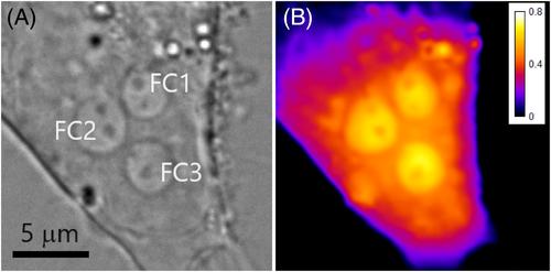
蛋白质浓度(PC)是细胞和细胞器的一个基本特征;它决定了大分子拥挤效应的程度,是细胞健康的一个敏感指标。基于明视野的强度传输方程(TIE)成像与体积测量相结合,为量化蛋白质浓度提供了一种简单而直接的方法。然而,由于 TIE 基于几何光学原理,其对微米大小颗粒的适用性尚不明确。在这里,我们展示了 TIE 可用于尺寸与波长相当的粒子。同时,我们还介绍了一种新的 ImageJ 插件,无需借助高级数学程序即可处理 TIE 图像。要将 TIE 数据转换为 PC,了解颗粒体积至关重要。细菌或其他孤立颗粒的体积可通过外部吸收染料的位移来测量("透射染料 "或 TTD 显微镜),而对于球形细胞内颗粒,体积可通过其直径来估算。我们举例说明了 TIE 在大肠杆菌、哺乳动物核小体和核小体纤维中心的应用。该方法简单易用,空间分辨率高。
Measurement of protein concentration in bacteria and small organelles under a light transmission microscope
Protein concentration (PC) is an essential characteristic of cells and organelles; it determines the extent of macromolecular crowding effects and serves as a sensitive indicator of cellular health. A simple and direct way to quantify PC is provided by brightfield-based transport-of-intensity equation (TIE) imaging combined with volume measurements. However, since TIE is based on geometric optics, its applicability to micrometer-sized particles is not clear. Here, we show that TIE can be used on particles with sizes comparable to the wavelength. At the same time, we introduce a new ImageJ plugin that allows TIE image processing without resorting to advanced mathematical programs. To convert TIE data to PC, knowledge of particle volumes is essential. The volumes of bacteria or other isolated particles can be measured by displacement of an external absorbing dye (“transmission-through-dye” or TTD microscopy), and for spherical intracellular particles, volumes can be estimated from their diameters. We illustrate the use of TIE on Escherichia coli, mammalian nucleoli, and nucleolar fibrillar centers. The method is easy to use and achieves high spatial resolution.
期刊介绍:
Journal of Molecular Recognition (JMR) publishes original research papers and reviews describing substantial advances in our understanding of molecular recognition phenomena in life sciences, covering all aspects from biochemistry, molecular biology, medicine, and biophysics. The research may employ experimental, theoretical and/or computational approaches.
The focus of the journal is on recognition phenomena involving biomolecules and their biological / biochemical partners rather than on the recognition of metal ions or inorganic compounds. Molecular recognition involves non-covalent specific interactions between two or more biological molecules, molecular aggregates, cellular modules or organelles, as exemplified by receptor-ligand, antigen-antibody, nucleic acid-protein, sugar-lectin, to mention just a few of the possible interactions. The journal invites manuscripts that aim to achieve a complete description of molecular recognition mechanisms between well-characterized biomolecules in terms of structure, dynamics and biological activity. Such studies may help the future development of new drugs and vaccines, although the experimental testing of new drugs and vaccines falls outside the scope of the journal. Manuscripts that describe the application of standard approaches and techniques to design or model new molecular entities or to describe interactions between biomolecules, but do not provide new insights into molecular recognition processes will not be considered. Similarly, manuscripts involving biomolecules uncharacterized at the sequence level (e.g. calf thymus DNA) will not be considered.
 求助内容:
求助内容: 应助结果提醒方式:


