Aditya D Pradana, Arditya Damarkusuma, Hariadi Hariawan
{"title":"步入光明:界定非 ST 段抬高型心肌梗死的罪魁祸首病变。","authors":"Aditya D Pradana, Arditya Damarkusuma, Hariadi Hariawan","doi":"10.37616/2212-5043.1377","DOIUrl":null,"url":null,"abstract":"<p><p>Identifying the infarct-related artery (IRA) in a non-ST-segment-elevation acute myocardial infarction (NSTEMI) can be very challenging, particularly in a hospital that cannot perform intracoronary imaging due to certain limitations. This is because, by angiography, most patients present with multivessel coronary artery disease (CAD), diffuse disease, or non-significant CAD. We present a case of a 60-year-old female patient presented with substernal chest pain and palpitations of 6 h duration. The first hospital contact 12-lead electrocardiogram (ECG) showed ventricular tachycardia (VT) with unstable hemodynamics, after stabilization patient was transported to the catheterization laboratory for immediate percutaneous coronary intervention (PCI). With a clue of VT morphology, post-converted ECG, and coronary angiography, the patient successfully underwent PCI in the left circumflex artery.</p>","PeriodicalId":17319,"journal":{"name":"Journal of the Saudi Heart Association","volume":"36 2","pages":"94-98"},"PeriodicalIF":1.3000,"publicationDate":"2024-06-10","publicationTypes":"Journal Article","fieldsOfStudy":null,"isOpenAccess":false,"openAccessPdf":"https://www.ncbi.nlm.nih.gov/pmc/articles/PMC11195660/pdf/","citationCount":"0","resultStr":"{\"title\":\"Stepping into the Light: Defining Culprit Lesion in Non-ST Elevation Myocardial Infarction.\",\"authors\":\"Aditya D Pradana, Arditya Damarkusuma, Hariadi Hariawan\",\"doi\":\"10.37616/2212-5043.1377\",\"DOIUrl\":null,\"url\":null,\"abstract\":\"<p><p>Identifying the infarct-related artery (IRA) in a non-ST-segment-elevation acute myocardial infarction (NSTEMI) can be very challenging, particularly in a hospital that cannot perform intracoronary imaging due to certain limitations. This is because, by angiography, most patients present with multivessel coronary artery disease (CAD), diffuse disease, or non-significant CAD. We present a case of a 60-year-old female patient presented with substernal chest pain and palpitations of 6 h duration. The first hospital contact 12-lead electrocardiogram (ECG) showed ventricular tachycardia (VT) with unstable hemodynamics, after stabilization patient was transported to the catheterization laboratory for immediate percutaneous coronary intervention (PCI). With a clue of VT morphology, post-converted ECG, and coronary angiography, the patient successfully underwent PCI in the left circumflex artery.</p>\",\"PeriodicalId\":17319,\"journal\":{\"name\":\"Journal of the Saudi Heart Association\",\"volume\":\"36 2\",\"pages\":\"94-98\"},\"PeriodicalIF\":1.3000,\"publicationDate\":\"2024-06-10\",\"publicationTypes\":\"Journal Article\",\"fieldsOfStudy\":null,\"isOpenAccess\":false,\"openAccessPdf\":\"https://www.ncbi.nlm.nih.gov/pmc/articles/PMC11195660/pdf/\",\"citationCount\":\"0\",\"resultStr\":null,\"platform\":\"Semanticscholar\",\"paperid\":null,\"PeriodicalName\":\"Journal of the Saudi Heart Association\",\"FirstCategoryId\":\"1085\",\"ListUrlMain\":\"https://doi.org/10.37616/2212-5043.1377\",\"RegionNum\":0,\"RegionCategory\":null,\"ArticlePicture\":[],\"TitleCN\":null,\"AbstractTextCN\":null,\"PMCID\":null,\"EPubDate\":\"2024/1/1 0:00:00\",\"PubModel\":\"eCollection\",\"JCR\":\"Q4\",\"JCRName\":\"CARDIAC & CARDIOVASCULAR SYSTEMS\",\"Score\":null,\"Total\":0}","platform":"Semanticscholar","paperid":null,"PeriodicalName":"Journal of the Saudi Heart Association","FirstCategoryId":"1085","ListUrlMain":"https://doi.org/10.37616/2212-5043.1377","RegionNum":0,"RegionCategory":null,"ArticlePicture":[],"TitleCN":null,"AbstractTextCN":null,"PMCID":null,"EPubDate":"2024/1/1 0:00:00","PubModel":"eCollection","JCR":"Q4","JCRName":"CARDIAC & CARDIOVASCULAR SYSTEMS","Score":null,"Total":0}
Stepping into the Light: Defining Culprit Lesion in Non-ST Elevation Myocardial Infarction.
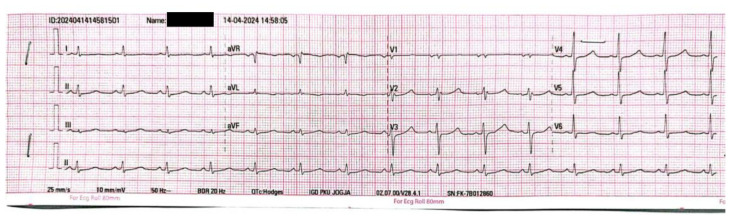
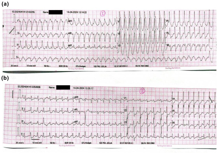
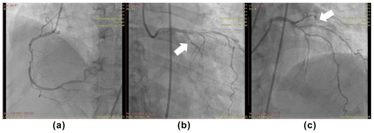
Identifying the infarct-related artery (IRA) in a non-ST-segment-elevation acute myocardial infarction (NSTEMI) can be very challenging, particularly in a hospital that cannot perform intracoronary imaging due to certain limitations. This is because, by angiography, most patients present with multivessel coronary artery disease (CAD), diffuse disease, or non-significant CAD. We present a case of a 60-year-old female patient presented with substernal chest pain and palpitations of 6 h duration. The first hospital contact 12-lead electrocardiogram (ECG) showed ventricular tachycardia (VT) with unstable hemodynamics, after stabilization patient was transported to the catheterization laboratory for immediate percutaneous coronary intervention (PCI). With a clue of VT morphology, post-converted ECG, and coronary angiography, the patient successfully underwent PCI in the left circumflex artery.

 求助内容:
求助内容: 应助结果提醒方式:


