Jin-Hui Yin, Ya-Ou Liu, Hong-Liang Li, Jean Marc Burgunder, Yue Huang
{"title":"通过扩散峰度和扩散张量成像揭示突变亨廷汀基因携带者的白质微结构变化","authors":"Jin-Hui Yin, Ya-Ou Liu, Hong-Liang Li, Jean Marc Burgunder, Yue Huang","doi":"10.3233/JHD-240018","DOIUrl":null,"url":null,"abstract":"<p><strong>Background: </strong>Diffusion magnetic resonance imaging (dMRI) has revealed microstructural changes in white matter (WM) in Huntington's disease (HD).</p><p><strong>Objective: </strong>To compare the validities of different dMRI, i.e., diffusion kurtosis imaging (DKI) and diffusion tensor imaging (DTI) in HD.</p><p><strong>Methods: </strong>22 mutant huntingtin (mHTT) carriers and 14 controls were enrolled. Clinical assessments and dMRI were conducted. Based on CAG-Age Product (CAP) score, mHTT carriers were categorized into high CAP (hCAP) and medium and low CAP (m& lCAP) groups. Spearman analyses were used to explore correlations between imaging parameters in brain regions and clinical assessments. Receiver operating characteristic (ROC) was used to distinguish mHTT carriers from control, and define the HD patients at advanced stage.</p><p><strong>Results: </strong>Compared to controls, mHTT carriers exhibited WM changes in DKI and DTI. There were 22 more regions showing significant differences in HD detected by MK than FA. Only MK in five brain regions showed significantly difference between any two group, and negatively correlated with the disease burden (r = -0.80 to -0.71). ROC analysis revealed that MK was more sensitive and FA was more specific, while Youden index showed that the integration of FA and MK gave rise to higher authenticities, in distinguishing m& lCAP from controls (Youden Index = 0.786), and discerning different phase of HD (Youden Index = 0.804).</p><p><strong>Conclusions: </strong>Microstructural changes in WM occur at early stage of HD and deteriorate over the disease progression. Integrating DKI and DTI would provide the best accuracies for differentiating early HD from control and identifying advanced HD.</p>","PeriodicalId":16042,"journal":{"name":"Journal of Huntington's disease","volume":" ","pages":"301-313"},"PeriodicalIF":3.1000,"publicationDate":"2024-01-01","publicationTypes":"Journal Article","fieldsOfStudy":null,"isOpenAccess":false,"openAccessPdf":"https://www.ncbi.nlm.nih.gov/pmc/articles/PMC11494636/pdf/","citationCount":"0","resultStr":"{\"title\":\"White Matter Microstructure Changes Revealed by Diffusion Kurtosis and Diffusion Tensor Imaging in Mutant Huntingtin Gene Carriers.\",\"authors\":\"Jin-Hui Yin, Ya-Ou Liu, Hong-Liang Li, Jean Marc Burgunder, Yue Huang\",\"doi\":\"10.3233/JHD-240018\",\"DOIUrl\":null,\"url\":null,\"abstract\":\"<p><strong>Background: </strong>Diffusion magnetic resonance imaging (dMRI) has revealed microstructural changes in white matter (WM) in Huntington's disease (HD).</p><p><strong>Objective: </strong>To compare the validities of different dMRI, i.e., diffusion kurtosis imaging (DKI) and diffusion tensor imaging (DTI) in HD.</p><p><strong>Methods: </strong>22 mutant huntingtin (mHTT) carriers and 14 controls were enrolled. Clinical assessments and dMRI were conducted. Based on CAG-Age Product (CAP) score, mHTT carriers were categorized into high CAP (hCAP) and medium and low CAP (m& lCAP) groups. Spearman analyses were used to explore correlations between imaging parameters in brain regions and clinical assessments. Receiver operating characteristic (ROC) was used to distinguish mHTT carriers from control, and define the HD patients at advanced stage.</p><p><strong>Results: </strong>Compared to controls, mHTT carriers exhibited WM changes in DKI and DTI. There were 22 more regions showing significant differences in HD detected by MK than FA. Only MK in five brain regions showed significantly difference between any two group, and negatively correlated with the disease burden (r = -0.80 to -0.71). ROC analysis revealed that MK was more sensitive and FA was more specific, while Youden index showed that the integration of FA and MK gave rise to higher authenticities, in distinguishing m& lCAP from controls (Youden Index = 0.786), and discerning different phase of HD (Youden Index = 0.804).</p><p><strong>Conclusions: </strong>Microstructural changes in WM occur at early stage of HD and deteriorate over the disease progression. Integrating DKI and DTI would provide the best accuracies for differentiating early HD from control and identifying advanced HD.</p>\",\"PeriodicalId\":16042,\"journal\":{\"name\":\"Journal of Huntington's disease\",\"volume\":\" \",\"pages\":\"301-313\"},\"PeriodicalIF\":3.1000,\"publicationDate\":\"2024-01-01\",\"publicationTypes\":\"Journal Article\",\"fieldsOfStudy\":null,\"isOpenAccess\":false,\"openAccessPdf\":\"https://www.ncbi.nlm.nih.gov/pmc/articles/PMC11494636/pdf/\",\"citationCount\":\"0\",\"resultStr\":null,\"platform\":\"Semanticscholar\",\"paperid\":null,\"PeriodicalName\":\"Journal of Huntington's disease\",\"FirstCategoryId\":\"1085\",\"ListUrlMain\":\"https://doi.org/10.3233/JHD-240018\",\"RegionNum\":0,\"RegionCategory\":null,\"ArticlePicture\":[],\"TitleCN\":null,\"AbstractTextCN\":null,\"PMCID\":null,\"EPubDate\":\"\",\"PubModel\":\"\",\"JCR\":\"Q3\",\"JCRName\":\"NEUROSCIENCES\",\"Score\":null,\"Total\":0}","platform":"Semanticscholar","paperid":null,"PeriodicalName":"Journal of Huntington's disease","FirstCategoryId":"1085","ListUrlMain":"https://doi.org/10.3233/JHD-240018","RegionNum":0,"RegionCategory":null,"ArticlePicture":[],"TitleCN":null,"AbstractTextCN":null,"PMCID":null,"EPubDate":"","PubModel":"","JCR":"Q3","JCRName":"NEUROSCIENCES","Score":null,"Total":0}
引用次数: 0
摘要
背景:扩散磁共振成像(dMRI)揭示了亨廷顿氏病(HD)白质(WM)的微观结构变化:比较不同 dMRI(即扩散峰度成像(DKI)和扩散张量成像(DTI))在 HD 中的有效性。进行了临床评估和 dMRI 检查。根据 CAG 年龄乘积(CAP)得分,将 mHTT 携带者分为高 CAP 组(hCAP)和中低 CAP 组(m& lCAP)。斯皮尔曼分析用于探讨脑区成像参数与临床评估之间的相关性。受体操作特征(ROC)用于区分 mHTT 携带者和对照组,并界定晚期 HD 患者:与对照组相比,mHTT 携带者在 DKI 和 DTI 中表现出 WM 变化。与FA相比,MK检测到的HD显著差异区域多出22个。只有 5 个脑区的 MK 在任何两个组别之间存在显著差异,且与疾病负担呈负相关(r = -0.80 至 -0.71)。ROC分析显示,MK更灵敏,FA更特异,而Youden指数显示,在区分m& lCAP与对照组(Youden指数=0.786)和鉴别HD的不同阶段(Youden指数=0.804)时,FA和MK的整合具有更高的真实性:结论:WM的微结构变化发生在HD的早期阶段,并随着疾病的进展而恶化。整合 DKI 和 DTI 将为区分早期 HD 和对照组以及识别晚期 HD 提供最佳准确性。
White Matter Microstructure Changes Revealed by Diffusion Kurtosis and Diffusion Tensor Imaging in Mutant Huntingtin Gene Carriers.
Background: Diffusion magnetic resonance imaging (dMRI) has revealed microstructural changes in white matter (WM) in Huntington's disease (HD).
Objective: To compare the validities of different dMRI, i.e., diffusion kurtosis imaging (DKI) and diffusion tensor imaging (DTI) in HD.
Methods: 22 mutant huntingtin (mHTT) carriers and 14 controls were enrolled. Clinical assessments and dMRI were conducted. Based on CAG-Age Product (CAP) score, mHTT carriers were categorized into high CAP (hCAP) and medium and low CAP (m& lCAP) groups. Spearman analyses were used to explore correlations between imaging parameters in brain regions and clinical assessments. Receiver operating characteristic (ROC) was used to distinguish mHTT carriers from control, and define the HD patients at advanced stage.
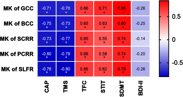
Results: Compared to controls, mHTT carriers exhibited WM changes in DKI and DTI. There were 22 more regions showing significant differences in HD detected by MK than FA. Only MK in five brain regions showed significantly difference between any two group, and negatively correlated with the disease burden (r = -0.80 to -0.71). ROC analysis revealed that MK was more sensitive and FA was more specific, while Youden index showed that the integration of FA and MK gave rise to higher authenticities, in distinguishing m& lCAP from controls (Youden Index = 0.786), and discerning different phase of HD (Youden Index = 0.804).
Conclusions: Microstructural changes in WM occur at early stage of HD and deteriorate over the disease progression. Integrating DKI and DTI would provide the best accuracies for differentiating early HD from control and identifying advanced HD.

 求助内容:
求助内容: 应助结果提醒方式:


