Per Thunswärd, Ellen Bergkvist, Liya Vishnevskaya, Yngve Forslin, Håkan Ahlström
{"title":"超声造影剂针引流:猪肝脏模型中超声活检针可见度的影响。","authors":"Per Thunswärd, Ellen Bergkvist, Liya Vishnevskaya, Yngve Forslin, Håkan Ahlström","doi":"10.1007/s00270-024-03758-1","DOIUrl":null,"url":null,"abstract":"<p><strong>Purpose: </strong>The visibility of biopsy needles in contrast-specific imaging mode can be improved by priming them with an ultrasound contrast agent (previously demonstrated in a phantom model/ex vivo). The purpose of this study was to validate this priming method in a porcine in vivo model.</p><p><strong>Materials and methods: </strong>Using a small syringe, full-core biopsy needles were primed with sulfur hexafluoride, an ultrasound contrast agent, with non-primed needles serving as controls (n = 30 + 30). Liver punctures were performed in a porcine model following intravenous administration of the same ultrasound contrast agent. Needle visibility, both in their entirety and at the tips, was evaluated in split-screen mode using contrast-specific imaging and B-mode (low mechanical index). The assessment included quantitative analysis, calculating the contrast-to-noise ratio, and qualitative evaluation through structured grading by three radiologists.</p><p><strong>Results: </strong>After needle priming, the contrast-to-noise ratio was superior for the needle in its entirety in contrast-specific imaging mode (p < 0.001) and slightly inferior in B-mode (p = 0.008). No differences were observed for the needle tips in either imaging mode. Qualitatively, the needle visibility was deemed clinically superior after needle priming throughout in contrast-specific imaging mode (p < 0.001), whereas no clinically relevant differences in B-mode for either the needle in its entirety (p = 0.11) or the needle tip (p = 1) were observed.</p><p><strong>Conclusion: </strong>In this in vivo porcine liver model experiment, priming biopsy needles with ultrasound contrast agent improved needle visibility in contrast-specific imaging mode but slightly reduced it in B-mode. These findings support the method's use for biopsies requiring target visualization in contrast-specific imaging mode.</p><p><strong>No level of evidence: </strong></p>","PeriodicalId":9591,"journal":{"name":"CardioVascular and Interventional Radiology","volume":null,"pages":null},"PeriodicalIF":2.8000,"publicationDate":"2024-07-01","publicationTypes":"Journal Article","fieldsOfStudy":null,"isOpenAccess":false,"openAccessPdf":"https://www.ncbi.nlm.nih.gov/pmc/articles/PMC11239778/pdf/","citationCount":"0","resultStr":"{\"title\":\"Ultrasound Contrast Agent Needle Priming: Impact on Sonographic Biopsy Needle Visibility in a Porcine Liver Model.\",\"authors\":\"Per Thunswärd, Ellen Bergkvist, Liya Vishnevskaya, Yngve Forslin, Håkan Ahlström\",\"doi\":\"10.1007/s00270-024-03758-1\",\"DOIUrl\":null,\"url\":null,\"abstract\":\"<p><strong>Purpose: </strong>The visibility of biopsy needles in contrast-specific imaging mode can be improved by priming them with an ultrasound contrast agent (previously demonstrated in a phantom model/ex vivo). The purpose of this study was to validate this priming method in a porcine in vivo model.</p><p><strong>Materials and methods: </strong>Using a small syringe, full-core biopsy needles were primed with sulfur hexafluoride, an ultrasound contrast agent, with non-primed needles serving as controls (n = 30 + 30). Liver punctures were performed in a porcine model following intravenous administration of the same ultrasound contrast agent. Needle visibility, both in their entirety and at the tips, was evaluated in split-screen mode using contrast-specific imaging and B-mode (low mechanical index). The assessment included quantitative analysis, calculating the contrast-to-noise ratio, and qualitative evaluation through structured grading by three radiologists.</p><p><strong>Results: </strong>After needle priming, the contrast-to-noise ratio was superior for the needle in its entirety in contrast-specific imaging mode (p < 0.001) and slightly inferior in B-mode (p = 0.008). No differences were observed for the needle tips in either imaging mode. Qualitatively, the needle visibility was deemed clinically superior after needle priming throughout in contrast-specific imaging mode (p < 0.001), whereas no clinically relevant differences in B-mode for either the needle in its entirety (p = 0.11) or the needle tip (p = 1) were observed.</p><p><strong>Conclusion: </strong>In this in vivo porcine liver model experiment, priming biopsy needles with ultrasound contrast agent improved needle visibility in contrast-specific imaging mode but slightly reduced it in B-mode. These findings support the method's use for biopsies requiring target visualization in contrast-specific imaging mode.</p><p><strong>No level of evidence: </strong></p>\",\"PeriodicalId\":9591,\"journal\":{\"name\":\"CardioVascular and Interventional Radiology\",\"volume\":null,\"pages\":null},\"PeriodicalIF\":2.8000,\"publicationDate\":\"2024-07-01\",\"publicationTypes\":\"Journal Article\",\"fieldsOfStudy\":null,\"isOpenAccess\":false,\"openAccessPdf\":\"https://www.ncbi.nlm.nih.gov/pmc/articles/PMC11239778/pdf/\",\"citationCount\":\"0\",\"resultStr\":null,\"platform\":\"Semanticscholar\",\"paperid\":null,\"PeriodicalName\":\"CardioVascular and Interventional Radiology\",\"FirstCategoryId\":\"3\",\"ListUrlMain\":\"https://doi.org/10.1007/s00270-024-03758-1\",\"RegionNum\":3,\"RegionCategory\":\"医学\",\"ArticlePicture\":[],\"TitleCN\":null,\"AbstractTextCN\":null,\"PMCID\":null,\"EPubDate\":\"2024/6/19 0:00:00\",\"PubModel\":\"Epub\",\"JCR\":\"Q2\",\"JCRName\":\"CARDIAC & CARDIOVASCULAR SYSTEMS\",\"Score\":null,\"Total\":0}","platform":"Semanticscholar","paperid":null,"PeriodicalName":"CardioVascular and Interventional Radiology","FirstCategoryId":"3","ListUrlMain":"https://doi.org/10.1007/s00270-024-03758-1","RegionNum":3,"RegionCategory":"医学","ArticlePicture":[],"TitleCN":null,"AbstractTextCN":null,"PMCID":null,"EPubDate":"2024/6/19 0:00:00","PubModel":"Epub","JCR":"Q2","JCRName":"CARDIAC & CARDIOVASCULAR SYSTEMS","Score":null,"Total":0}
引用次数: 0
摘要
目的:活检针在造影剂特异性成像模式下的可见度可通过使用超声造影剂来提高(之前已在模型/体内外进行过验证)。本研究的目的是在猪体内模型中验证这种引导方法:使用小型注射器,用超声造影剂六氟化硫为全芯活检针打底,未打底的针作为对照组(n = 30 + 30)。静脉注射相同的超声造影剂后,在猪模型中进行肝穿刺。使用对比度特异性成像和 B 模式(低机械指数),在分屏模式下对针头整体和针尖的可见度进行评估。评估包括定量分析、计算对比度与噪声比,以及由三位放射科医生进行结构化分级的定性评估:结果:在针引流后,造影剂特异性成像模式下,针整体的对比度与噪声比更优(P 结论:针引流后,造影剂特异性成像模式下,针整体的对比度与噪声比更优:在这个活体猪肝模型实验中,用超声造影剂引导活检针提高了造影剂特异性成像模式下的针头能见度,但在 B 模式下略微降低了能见度。这些发现支持将该方法用于需要在造影剂特异性成像模式下观察目标的活检:
Ultrasound Contrast Agent Needle Priming: Impact on Sonographic Biopsy Needle Visibility in a Porcine Liver Model.
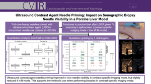
Purpose: The visibility of biopsy needles in contrast-specific imaging mode can be improved by priming them with an ultrasound contrast agent (previously demonstrated in a phantom model/ex vivo). The purpose of this study was to validate this priming method in a porcine in vivo model.
Materials and methods: Using a small syringe, full-core biopsy needles were primed with sulfur hexafluoride, an ultrasound contrast agent, with non-primed needles serving as controls (n = 30 + 30). Liver punctures were performed in a porcine model following intravenous administration of the same ultrasound contrast agent. Needle visibility, both in their entirety and at the tips, was evaluated in split-screen mode using contrast-specific imaging and B-mode (low mechanical index). The assessment included quantitative analysis, calculating the contrast-to-noise ratio, and qualitative evaluation through structured grading by three radiologists.
Results: After needle priming, the contrast-to-noise ratio was superior for the needle in its entirety in contrast-specific imaging mode (p < 0.001) and slightly inferior in B-mode (p = 0.008). No differences were observed for the needle tips in either imaging mode. Qualitatively, the needle visibility was deemed clinically superior after needle priming throughout in contrast-specific imaging mode (p < 0.001), whereas no clinically relevant differences in B-mode for either the needle in its entirety (p = 0.11) or the needle tip (p = 1) were observed.
Conclusion: In this in vivo porcine liver model experiment, priming biopsy needles with ultrasound contrast agent improved needle visibility in contrast-specific imaging mode but slightly reduced it in B-mode. These findings support the method's use for biopsies requiring target visualization in contrast-specific imaging mode.
期刊介绍:
CardioVascular and Interventional Radiology (CVIR) is the official journal of the Cardiovascular and Interventional Radiological Society of Europe, and is also the official organ of a number of additional distinguished national and international interventional radiological societies. CVIR publishes double blinded peer-reviewed original research work including clinical and laboratory investigations, technical notes, case reports, works in progress, and letters to the editor, as well as review articles, pictorial essays, editorials, and special invited submissions in the field of vascular and interventional radiology. Beside the communication of the latest research results in this field, it is also the aim of CVIR to support continuous medical education. Articles that are accepted for publication are done so with the understanding that they, or their substantive contents, have not been and will not be submitted to any other publication.
 求助内容:
求助内容: 应助结果提醒方式:


