{"title":"超越死亡:揭开细胞凋亡逃逸的复杂面纱。","authors":"Sercan Ergün, Senanur Aslan, Dilbeste Demir, Sümeyye Kayaoğlu, Mevsim Saydam, Yeda Keleş, Damla Kolcuoğlu, Neslihan Taşkurt Hekim, Sezgin Güneş","doi":"10.1007/s40291-024-00718-w","DOIUrl":null,"url":null,"abstract":"<p><p>Apoptosis, or programmed cell death, maintains tissue homeostasis by eliminating damaged or unnecessary cells. However, cells can evade this process, contributing to conditions such as cancer. Escape mechanisms include anoikis, mitochondrial DNA depletion, cellular FLICE inhibitory protein (c-FLIP), endosomal sorting complexes required for transport (ESCRT), mitotic slippage, anastasis, and blebbishield formation. Anoikis, triggered by cell detachment from the extracellular matrix, is pivotal in cancer research due to its role in cellular survival and metastasis. Mitochondrial DNA depletion, associated with cellular dysfunction and diseases such as breast and prostate cancer, links to apoptosis resistance. The c-FLIP protein family, notably CFLAR, regulates cell death processes as a truncated caspase-8 form. The ESCRT complex aids apoptosis evasion by repairing intracellular damage through increased Ca2+ levels. Antimitotic agents induce mitotic arrest in cancer treatment but can lead to mitotic slippage and tetraploid cell formation. Anastasis allows cells to resist apoptosis induced by various triggers. Blebbishield formation suppresses apoptosis indirectly in cancer stem cells by transforming apoptotic cells into blebbishields. In conclusion, the future of apoptosis research offers exciting possibilities for innovative therapeutic approaches, enhanced diagnostic tools, and a deeper understanding of the complex biological processes that govern cell fate. Collaborative efforts across disciplines, including molecular biology, genetics, immunology, and bioinformatics, will be essential to realize these prospects and improve patient outcomes in diverse disease contexts.</p>","PeriodicalId":49797,"journal":{"name":"Molecular Diagnosis & Therapy","volume":" ","pages":"403-423"},"PeriodicalIF":4.4000,"publicationDate":"2024-07-01","publicationTypes":"Journal Article","fieldsOfStudy":null,"isOpenAccess":false,"openAccessPdf":"https://www.ncbi.nlm.nih.gov/pmc/articles/PMC11211167/pdf/","citationCount":"0","resultStr":"{\"title\":\"Beyond Death: Unmasking the Intricacies of Apoptosis Escape.\",\"authors\":\"Sercan Ergün, Senanur Aslan, Dilbeste Demir, Sümeyye Kayaoğlu, Mevsim Saydam, Yeda Keleş, Damla Kolcuoğlu, Neslihan Taşkurt Hekim, Sezgin Güneş\",\"doi\":\"10.1007/s40291-024-00718-w\",\"DOIUrl\":null,\"url\":null,\"abstract\":\"<p><p>Apoptosis, or programmed cell death, maintains tissue homeostasis by eliminating damaged or unnecessary cells. However, cells can evade this process, contributing to conditions such as cancer. Escape mechanisms include anoikis, mitochondrial DNA depletion, cellular FLICE inhibitory protein (c-FLIP), endosomal sorting complexes required for transport (ESCRT), mitotic slippage, anastasis, and blebbishield formation. Anoikis, triggered by cell detachment from the extracellular matrix, is pivotal in cancer research due to its role in cellular survival and metastasis. Mitochondrial DNA depletion, associated with cellular dysfunction and diseases such as breast and prostate cancer, links to apoptosis resistance. The c-FLIP protein family, notably CFLAR, regulates cell death processes as a truncated caspase-8 form. The ESCRT complex aids apoptosis evasion by repairing intracellular damage through increased Ca2+ levels. Antimitotic agents induce mitotic arrest in cancer treatment but can lead to mitotic slippage and tetraploid cell formation. Anastasis allows cells to resist apoptosis induced by various triggers. Blebbishield formation suppresses apoptosis indirectly in cancer stem cells by transforming apoptotic cells into blebbishields. In conclusion, the future of apoptosis research offers exciting possibilities for innovative therapeutic approaches, enhanced diagnostic tools, and a deeper understanding of the complex biological processes that govern cell fate. Collaborative efforts across disciplines, including molecular biology, genetics, immunology, and bioinformatics, will be essential to realize these prospects and improve patient outcomes in diverse disease contexts.</p>\",\"PeriodicalId\":49797,\"journal\":{\"name\":\"Molecular Diagnosis & Therapy\",\"volume\":\" \",\"pages\":\"403-423\"},\"PeriodicalIF\":4.4000,\"publicationDate\":\"2024-07-01\",\"publicationTypes\":\"Journal Article\",\"fieldsOfStudy\":null,\"isOpenAccess\":false,\"openAccessPdf\":\"https://www.ncbi.nlm.nih.gov/pmc/articles/PMC11211167/pdf/\",\"citationCount\":\"0\",\"resultStr\":null,\"platform\":\"Semanticscholar\",\"paperid\":null,\"PeriodicalName\":\"Molecular Diagnosis & Therapy\",\"FirstCategoryId\":\"3\",\"ListUrlMain\":\"https://doi.org/10.1007/s40291-024-00718-w\",\"RegionNum\":3,\"RegionCategory\":\"医学\",\"ArticlePicture\":[],\"TitleCN\":null,\"AbstractTextCN\":null,\"PMCID\":null,\"EPubDate\":\"2024/6/18 0:00:00\",\"PubModel\":\"Epub\",\"JCR\":\"Q1\",\"JCRName\":\"GENETICS & HEREDITY\",\"Score\":null,\"Total\":0}","platform":"Semanticscholar","paperid":null,"PeriodicalName":"Molecular Diagnosis & Therapy","FirstCategoryId":"3","ListUrlMain":"https://doi.org/10.1007/s40291-024-00718-w","RegionNum":3,"RegionCategory":"医学","ArticlePicture":[],"TitleCN":null,"AbstractTextCN":null,"PMCID":null,"EPubDate":"2024/6/18 0:00:00","PubModel":"Epub","JCR":"Q1","JCRName":"GENETICS & HEREDITY","Score":null,"Total":0}
引用次数: 0
摘要
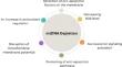
细胞凋亡(或称程序性细胞死亡)通过清除受损或不必要的细胞来维持组织的平衡。然而,细胞可以逃避这一过程,从而导致癌症等病症。细胞凋亡的逃逸机制包括anoikis、线粒体DNA耗竭、细胞FLICE抑制蛋白(c-FLIP)、运输所需的内质体分拣复合物(ESCRT)、有丝分裂滑动、吻合和blebbishield形成。由细胞脱离细胞外基质引发的吻合作用在癌症研究中至关重要,因为它在细胞存活和转移中起着重要作用。线粒体 DNA 的耗竭与细胞功能障碍以及乳腺癌和前列腺癌等疾病有关,与细胞凋亡抵抗有关。c-FLIP 蛋白家族,特别是 CFLAR,以截短的 caspase-8 形式调节细胞死亡过程。ESCRT 复合物通过增加 Ca2+ 水平修复细胞内损伤,从而帮助逃避细胞凋亡。在癌症治疗中,抗有丝分裂药物可诱导有丝分裂停止,但也可能导致有丝分裂滑动和四倍体细胞的形成。吻合使细胞能够抵御各种诱因引起的细胞凋亡。通过将凋亡细胞转化为blebbishield,Blebbishield的形成间接抑制了癌症干细胞的凋亡。总之,细胞凋亡研究的未来为创新治疗方法、增强诊断工具以及深入了解支配细胞命运的复杂生物过程提供了令人兴奋的可能性。包括分子生物学、遗传学、免疫学和生物信息学在内的跨学科合作对于实现这些前景和改善不同疾病背景下的患者预后至关重要。
Beyond Death: Unmasking the Intricacies of Apoptosis Escape.
Apoptosis, or programmed cell death, maintains tissue homeostasis by eliminating damaged or unnecessary cells. However, cells can evade this process, contributing to conditions such as cancer. Escape mechanisms include anoikis, mitochondrial DNA depletion, cellular FLICE inhibitory protein (c-FLIP), endosomal sorting complexes required for transport (ESCRT), mitotic slippage, anastasis, and blebbishield formation. Anoikis, triggered by cell detachment from the extracellular matrix, is pivotal in cancer research due to its role in cellular survival and metastasis. Mitochondrial DNA depletion, associated with cellular dysfunction and diseases such as breast and prostate cancer, links to apoptosis resistance. The c-FLIP protein family, notably CFLAR, regulates cell death processes as a truncated caspase-8 form. The ESCRT complex aids apoptosis evasion by repairing intracellular damage through increased Ca2+ levels. Antimitotic agents induce mitotic arrest in cancer treatment but can lead to mitotic slippage and tetraploid cell formation. Anastasis allows cells to resist apoptosis induced by various triggers. Blebbishield formation suppresses apoptosis indirectly in cancer stem cells by transforming apoptotic cells into blebbishields. In conclusion, the future of apoptosis research offers exciting possibilities for innovative therapeutic approaches, enhanced diagnostic tools, and a deeper understanding of the complex biological processes that govern cell fate. Collaborative efforts across disciplines, including molecular biology, genetics, immunology, and bioinformatics, will be essential to realize these prospects and improve patient outcomes in diverse disease contexts.
期刊介绍:
Molecular Diagnosis & Therapy welcomes current opinion articles on emerging or contentious issues, comprehensive narrative reviews, systematic reviews (as outlined by the PRISMA statement), original research articles (including short communications) and letters to the editor. All manuscripts are subject to peer review by international experts.
 求助内容:
求助内容: 应助结果提醒方式:


