{"title":"虹膜内皮综合征患者的体内共焦显微镜和前眼球光学相干断层扫描结果。","authors":"Gülay Güler Canözer, Emine Tınkır Kayıtmazbatır, Esra Öztürk, Ayşe Bozkurt Oflaz, Banu Bozkurt","doi":"10.4274/tjo.galenos.2024.78861","DOIUrl":null,"url":null,"abstract":"<p><p>This case report aims to present the findings of in vivo confocal microscopy (IVCM) and anterior segment optical coherence tomography (AS-OCT) in three patients with iridocorneal endothelial (ICE) syndrome. Three female patients 37, 50, and 57 years of age presented with complaints of unilateral visual impairment and elevated intraocular pressure (IOP). Biomicroscopy revealed unilateral pupil irregularities and anterior synechiae, and gonioscopy demonstrated synechiae in the iridocorneal angle. IOP was within normal limits with medical treatment in two patients, while one patient had an IOP of 44 mmHg despite maximal antiglaucomatous treatment. IVCM revealed large, polymorphic, and hyperreflective cells in the corneal endothelial layer of the affected eyes and normal corneal epithelium, stroma, and endothelium in the fellow eyes. AS-OCT findings were normal in healthy eyes, while the affected eye showed synechiae in the iridocorneal angle and a hyperreflective, thickened endothelial layer. The patient with refractory glaucoma underwent trabeculectomy surgery with 5-fluorouracil. In conclusion, IVCM and AS-OCT allow a detailed examination of endothelial cell abnormalities and iridocorneal membranes in ICE syndrome, which is characterized by unilateral pupil and iris irregularities and anterior synechiae mainly in women.</p>","PeriodicalId":23373,"journal":{"name":"Turkish Journal of Ophthalmology","volume":" ","pages":"170-174"},"PeriodicalIF":0.0000,"publicationDate":"2024-06-28","publicationTypes":"Journal Article","fieldsOfStudy":null,"isOpenAccess":false,"openAccessPdf":"https://www.ncbi.nlm.nih.gov/pmc/articles/PMC11589315/pdf/","citationCount":"0","resultStr":"{\"title\":\"<i>In Vivo</i> Confocal Microscopy and Anterior Segment Optical Coherence Tomography Findings of Patients with Iridocorneal Endothelial Syndrome\",\"authors\":\"Gülay Güler Canözer, Emine Tınkır Kayıtmazbatır, Esra Öztürk, Ayşe Bozkurt Oflaz, Banu Bozkurt\",\"doi\":\"10.4274/tjo.galenos.2024.78861\",\"DOIUrl\":null,\"url\":null,\"abstract\":\"<p><p>This case report aims to present the findings of in vivo confocal microscopy (IVCM) and anterior segment optical coherence tomography (AS-OCT) in three patients with iridocorneal endothelial (ICE) syndrome. Three female patients 37, 50, and 57 years of age presented with complaints of unilateral visual impairment and elevated intraocular pressure (IOP). Biomicroscopy revealed unilateral pupil irregularities and anterior synechiae, and gonioscopy demonstrated synechiae in the iridocorneal angle. IOP was within normal limits with medical treatment in two patients, while one patient had an IOP of 44 mmHg despite maximal antiglaucomatous treatment. IVCM revealed large, polymorphic, and hyperreflective cells in the corneal endothelial layer of the affected eyes and normal corneal epithelium, stroma, and endothelium in the fellow eyes. AS-OCT findings were normal in healthy eyes, while the affected eye showed synechiae in the iridocorneal angle and a hyperreflective, thickened endothelial layer. The patient with refractory glaucoma underwent trabeculectomy surgery with 5-fluorouracil. In conclusion, IVCM and AS-OCT allow a detailed examination of endothelial cell abnormalities and iridocorneal membranes in ICE syndrome, which is characterized by unilateral pupil and iris irregularities and anterior synechiae mainly in women.</p>\",\"PeriodicalId\":23373,\"journal\":{\"name\":\"Turkish Journal of Ophthalmology\",\"volume\":\" \",\"pages\":\"170-174\"},\"PeriodicalIF\":0.0000,\"publicationDate\":\"2024-06-28\",\"publicationTypes\":\"Journal Article\",\"fieldsOfStudy\":null,\"isOpenAccess\":false,\"openAccessPdf\":\"https://www.ncbi.nlm.nih.gov/pmc/articles/PMC11589315/pdf/\",\"citationCount\":\"0\",\"resultStr\":null,\"platform\":\"Semanticscholar\",\"paperid\":null,\"PeriodicalName\":\"Turkish Journal of Ophthalmology\",\"FirstCategoryId\":\"1085\",\"ListUrlMain\":\"https://doi.org/10.4274/tjo.galenos.2024.78861\",\"RegionNum\":0,\"RegionCategory\":null,\"ArticlePicture\":[],\"TitleCN\":null,\"AbstractTextCN\":null,\"PMCID\":null,\"EPubDate\":\"2024/6/12 0:00:00\",\"PubModel\":\"Epub\",\"JCR\":\"Q3\",\"JCRName\":\"Medicine\",\"Score\":null,\"Total\":0}","platform":"Semanticscholar","paperid":null,"PeriodicalName":"Turkish Journal of Ophthalmology","FirstCategoryId":"1085","ListUrlMain":"https://doi.org/10.4274/tjo.galenos.2024.78861","RegionNum":0,"RegionCategory":null,"ArticlePicture":[],"TitleCN":null,"AbstractTextCN":null,"PMCID":null,"EPubDate":"2024/6/12 0:00:00","PubModel":"Epub","JCR":"Q3","JCRName":"Medicine","Score":null,"Total":0}
In Vivo Confocal Microscopy and Anterior Segment Optical Coherence Tomography Findings of Patients with Iridocorneal Endothelial Syndrome
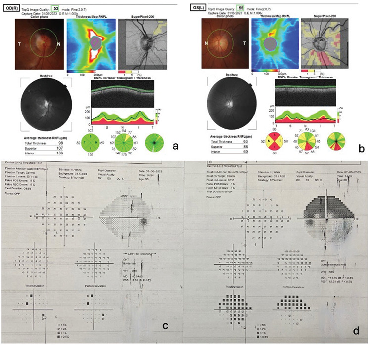
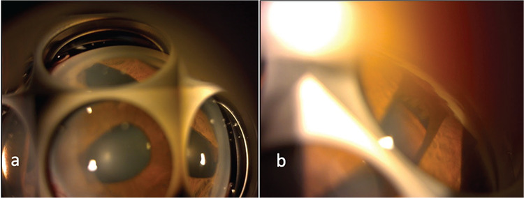
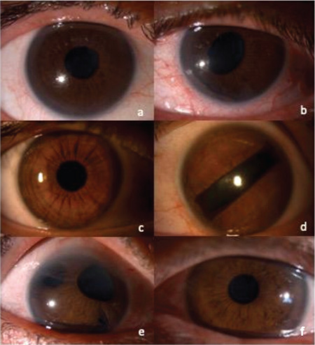
This case report aims to present the findings of in vivo confocal microscopy (IVCM) and anterior segment optical coherence tomography (AS-OCT) in three patients with iridocorneal endothelial (ICE) syndrome. Three female patients 37, 50, and 57 years of age presented with complaints of unilateral visual impairment and elevated intraocular pressure (IOP). Biomicroscopy revealed unilateral pupil irregularities and anterior synechiae, and gonioscopy demonstrated synechiae in the iridocorneal angle. IOP was within normal limits with medical treatment in two patients, while one patient had an IOP of 44 mmHg despite maximal antiglaucomatous treatment. IVCM revealed large, polymorphic, and hyperreflective cells in the corneal endothelial layer of the affected eyes and normal corneal epithelium, stroma, and endothelium in the fellow eyes. AS-OCT findings were normal in healthy eyes, while the affected eye showed synechiae in the iridocorneal angle and a hyperreflective, thickened endothelial layer. The patient with refractory glaucoma underwent trabeculectomy surgery with 5-fluorouracil. In conclusion, IVCM and AS-OCT allow a detailed examination of endothelial cell abnormalities and iridocorneal membranes in ICE syndrome, which is characterized by unilateral pupil and iris irregularities and anterior synechiae mainly in women.
期刊介绍:
The Turkish Journal of Ophthalmology (TJO) is the only scientific periodical publication of the Turkish Ophthalmological Association and has been published since January 1929. In its early years, the journal was published in Turkish and French. Although there were temporary interruptions in the publication of the journal due to various challenges, the Turkish Journal of Ophthalmology has been published continually from 1971 to the present. The target audience includes specialists and physicians in training in ophthalmology in all relevant disciplines.

 求助内容:
求助内容: 应助结果提醒方式:


