Lukas Mohl, Roger Karl, Matthias N Hagedorn, Armin Runz, Stephan Skornitzke, Malte Toelle, C Soeren Bergt, Johannes Hatzl, Christian Uhl, Dittmar Böckler, Katrin Meisenbacher, Sandy Engelhardt
{"title":"在 B 型主动脉夹层患者特异性灌注模型中模拟胸腔内血管主动脉修复术。","authors":"Lukas Mohl, Roger Karl, Matthias N Hagedorn, Armin Runz, Stephan Skornitzke, Malte Toelle, C Soeren Bergt, Johannes Hatzl, Christian Uhl, Dittmar Böckler, Katrin Meisenbacher, Sandy Engelhardt","doi":"10.1007/s11548-024-03190-3","DOIUrl":null,"url":null,"abstract":"<p><strong>Purpose: </strong>Complicated type B Aortic dissection is a severe aortic pathology that requires treatment through thoracic endovascular aortic repair (TEVAR). During TEVAR a stentgraft is deployed in the aortic lumen in order to restore blood flow. Due to the complicated pathology including an entry, a resulting dissection wall with potentially several re-entries, replicating this structure artificially has proven to be challenging thus far.</p><p><strong>Methods: </strong>We developed a 3d printed, patient-specific and perfused aortic dissection phantom with a flexible dissection flap and all major branching vessels. The model was segmented from CTA images and fabricated out of a flexible material to mimic aortic wall tissue. It was placed in a pulsatile hemodynamic flow loop. Hemodynamics were investigated through pressure and flow measurements and doppler ultrasound imaging. Surgeons performed a TEVAR intervention including stentgraft deployment under fluoroscopic guidance.</p><p><strong>Results: </strong>The flexible aortic dissection phantom was successfully incorporated in the hemodynamic flow loop, a systolic pressure of 112 mmHg and physiological flow of 4.05 L per minute was reached. Flow velocities were higher in true lumen with a up to 35.7 cm/s compared to the false lumen with a maximum of 13.3 cm/s, chaotic flow patterns were observed on main entry and reentry sights. A TEVAR procedure was successfully performed under fluoroscopy. The position of the stentgraft was confirmed using CTA imaging.</p><p><strong>Conclusions: </strong>This perfused in-vitro phantom allows for detailed investigation of the complex inner hemodynamics of aortic dissections on a patient-specific level and enables the simulation of TEVAR procedures in a real endovascular operating environment. Therefore, it could provide a dynamic platform for future surgical training and research.</p>","PeriodicalId":51251,"journal":{"name":"International Journal of Computer Assisted Radiology and Surgery","volume":" ","pages":"391-404"},"PeriodicalIF":2.3000,"publicationDate":"2025-02-01","publicationTypes":"Journal Article","fieldsOfStudy":null,"isOpenAccess":false,"openAccessPdf":"https://www.ncbi.nlm.nih.gov/pmc/articles/PMC11807923/pdf/","citationCount":"0","resultStr":"{\"title\":\"Simulation of thoracic endovascular aortic repair in a perfused patient-specific model of type B aortic dissection.\",\"authors\":\"Lukas Mohl, Roger Karl, Matthias N Hagedorn, Armin Runz, Stephan Skornitzke, Malte Toelle, C Soeren Bergt, Johannes Hatzl, Christian Uhl, Dittmar Böckler, Katrin Meisenbacher, Sandy Engelhardt\",\"doi\":\"10.1007/s11548-024-03190-3\",\"DOIUrl\":null,\"url\":null,\"abstract\":\"<p><strong>Purpose: </strong>Complicated type B Aortic dissection is a severe aortic pathology that requires treatment through thoracic endovascular aortic repair (TEVAR). During TEVAR a stentgraft is deployed in the aortic lumen in order to restore blood flow. Due to the complicated pathology including an entry, a resulting dissection wall with potentially several re-entries, replicating this structure artificially has proven to be challenging thus far.</p><p><strong>Methods: </strong>We developed a 3d printed, patient-specific and perfused aortic dissection phantom with a flexible dissection flap and all major branching vessels. The model was segmented from CTA images and fabricated out of a flexible material to mimic aortic wall tissue. It was placed in a pulsatile hemodynamic flow loop. Hemodynamics were investigated through pressure and flow measurements and doppler ultrasound imaging. Surgeons performed a TEVAR intervention including stentgraft deployment under fluoroscopic guidance.</p><p><strong>Results: </strong>The flexible aortic dissection phantom was successfully incorporated in the hemodynamic flow loop, a systolic pressure of 112 mmHg and physiological flow of 4.05 L per minute was reached. Flow velocities were higher in true lumen with a up to 35.7 cm/s compared to the false lumen with a maximum of 13.3 cm/s, chaotic flow patterns were observed on main entry and reentry sights. A TEVAR procedure was successfully performed under fluoroscopy. The position of the stentgraft was confirmed using CTA imaging.</p><p><strong>Conclusions: </strong>This perfused in-vitro phantom allows for detailed investigation of the complex inner hemodynamics of aortic dissections on a patient-specific level and enables the simulation of TEVAR procedures in a real endovascular operating environment. Therefore, it could provide a dynamic platform for future surgical training and research.</p>\",\"PeriodicalId\":51251,\"journal\":{\"name\":\"International Journal of Computer Assisted Radiology and Surgery\",\"volume\":\" \",\"pages\":\"391-404\"},\"PeriodicalIF\":2.3000,\"publicationDate\":\"2025-02-01\",\"publicationTypes\":\"Journal Article\",\"fieldsOfStudy\":null,\"isOpenAccess\":false,\"openAccessPdf\":\"https://www.ncbi.nlm.nih.gov/pmc/articles/PMC11807923/pdf/\",\"citationCount\":\"0\",\"resultStr\":null,\"platform\":\"Semanticscholar\",\"paperid\":null,\"PeriodicalName\":\"International Journal of Computer Assisted Radiology and Surgery\",\"FirstCategoryId\":\"5\",\"ListUrlMain\":\"https://doi.org/10.1007/s11548-024-03190-3\",\"RegionNum\":3,\"RegionCategory\":\"医学\",\"ArticlePicture\":[],\"TitleCN\":null,\"AbstractTextCN\":null,\"PMCID\":null,\"EPubDate\":\"2024/6/7 0:00:00\",\"PubModel\":\"Epub\",\"JCR\":\"Q3\",\"JCRName\":\"ENGINEERING, BIOMEDICAL\",\"Score\":null,\"Total\":0}","platform":"Semanticscholar","paperid":null,"PeriodicalName":"International Journal of Computer Assisted Radiology and Surgery","FirstCategoryId":"5","ListUrlMain":"https://doi.org/10.1007/s11548-024-03190-3","RegionNum":3,"RegionCategory":"医学","ArticlePicture":[],"TitleCN":null,"AbstractTextCN":null,"PMCID":null,"EPubDate":"2024/6/7 0:00:00","PubModel":"Epub","JCR":"Q3","JCRName":"ENGINEERING, BIOMEDICAL","Score":null,"Total":0}
Simulation of thoracic endovascular aortic repair in a perfused patient-specific model of type B aortic dissection.
Purpose: Complicated type B Aortic dissection is a severe aortic pathology that requires treatment through thoracic endovascular aortic repair (TEVAR). During TEVAR a stentgraft is deployed in the aortic lumen in order to restore blood flow. Due to the complicated pathology including an entry, a resulting dissection wall with potentially several re-entries, replicating this structure artificially has proven to be challenging thus far.
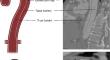
Methods: We developed a 3d printed, patient-specific and perfused aortic dissection phantom with a flexible dissection flap and all major branching vessels. The model was segmented from CTA images and fabricated out of a flexible material to mimic aortic wall tissue. It was placed in a pulsatile hemodynamic flow loop. Hemodynamics were investigated through pressure and flow measurements and doppler ultrasound imaging. Surgeons performed a TEVAR intervention including stentgraft deployment under fluoroscopic guidance.
Results: The flexible aortic dissection phantom was successfully incorporated in the hemodynamic flow loop, a systolic pressure of 112 mmHg and physiological flow of 4.05 L per minute was reached. Flow velocities were higher in true lumen with a up to 35.7 cm/s compared to the false lumen with a maximum of 13.3 cm/s, chaotic flow patterns were observed on main entry and reentry sights. A TEVAR procedure was successfully performed under fluoroscopy. The position of the stentgraft was confirmed using CTA imaging.
Conclusions: This perfused in-vitro phantom allows for detailed investigation of the complex inner hemodynamics of aortic dissections on a patient-specific level and enables the simulation of TEVAR procedures in a real endovascular operating environment. Therefore, it could provide a dynamic platform for future surgical training and research.
期刊介绍:
The International Journal for Computer Assisted Radiology and Surgery (IJCARS) is a peer-reviewed journal that provides a platform for closing the gap between medical and technical disciplines, and encourages interdisciplinary research and development activities in an international environment.

 求助内容:
求助内容: 应助结果提醒方式:


