{"title":"垂体腺瘤手术后的视觉通路恢复:视网膜结构、血管密度和神经传导分析的启示。","authors":"Yanhua Pang, Quanwen Zhao, Zeguang Huang, Kailun Lu, Fengyan Zhou, Wei Mo, Qianshuo Zhong, Zhi Tan","doi":"10.1007/s40123-024-00966-3","DOIUrl":null,"url":null,"abstract":"<p><strong>Introduction: </strong>This study investigates how surgery for pituitary adenoma (PA) affects the visual pathway, examining changes in the retina, blood vessel density, and nerve function. Since PAs often impair vision as a result of their location near visual structures, this research is key to understanding and improving vision recovery after surgery.</p><p><strong>Methods: </strong>Our study is based on a retrospective analysis of the historical data of 28 patients diagnosed with pituitary adenomas. We conducted assessments by reviewing preoperative and postoperative imaging records. These included optical coherence tomography (OCT) for retinal structure analysis, diffusion tensor imaging (DTI) for neural transmission evaluation, and optical coherence tomography angiography for assessing blood vessel density. These tools allowed for a detailed understanding of the structural and functional changes within the visual pathway following PA surgery.</p><p><strong>Results: </strong>OCT findings show postoperative changes in the eye: thinning in average and nasal circumpapillary retinal nerve fiber layer, thickening in macular central 1 mm inner plexus layer, ganglion cell complex, and nasal retinal nerve fiber layer. DTI reveals increased fractional anisotropy (FA) in the left optic chiasm and posterior optic nerve, decreased mid-segment optic nerve FA, and increased apparent diffusion coefficient (ADC) in the right optic chiasm and nerve segments. Early postoperative reduction in radial peripapillary capillaries plexus density is noted. Preoperative ganglion cell layer (GCL) thickness correlates with postoperative visual radiation FA and ADC values, especially in the inferior quadrant. A negative correlation exists between preoperative GCL thickness and postoperative visual field mean defect values, particularly on the temporal side and superior inner ring. All changes are statistically significant (P < 0.05).</p><p><strong>Conclusions: </strong>The study finds that surgery for PA has varied effects on vision. Early post surgery, there are changes in the retina and nerve signals. Macular GCL thickness before surgery might predict early visual recovery, influencing future research and treatment for vision issues related to PA.</p>","PeriodicalId":19623,"journal":{"name":"Ophthalmology and Therapy","volume":null,"pages":null},"PeriodicalIF":2.6000,"publicationDate":"2024-07-01","publicationTypes":"Journal Article","fieldsOfStudy":null,"isOpenAccess":false,"openAccessPdf":"https://www.ncbi.nlm.nih.gov/pmc/articles/PMC11178691/pdf/","citationCount":"0","resultStr":"{\"title\":\"Visual Pathway Recovery Post Pituitary Adenoma Surgery: Insights from Retinal Structure, Vascular Density, and Neural Conduction Analysis.\",\"authors\":\"Yanhua Pang, Quanwen Zhao, Zeguang Huang, Kailun Lu, Fengyan Zhou, Wei Mo, Qianshuo Zhong, Zhi Tan\",\"doi\":\"10.1007/s40123-024-00966-3\",\"DOIUrl\":null,\"url\":null,\"abstract\":\"<p><strong>Introduction: </strong>This study investigates how surgery for pituitary adenoma (PA) affects the visual pathway, examining changes in the retina, blood vessel density, and nerve function. Since PAs often impair vision as a result of their location near visual structures, this research is key to understanding and improving vision recovery after surgery.</p><p><strong>Methods: </strong>Our study is based on a retrospective analysis of the historical data of 28 patients diagnosed with pituitary adenomas. We conducted assessments by reviewing preoperative and postoperative imaging records. These included optical coherence tomography (OCT) for retinal structure analysis, diffusion tensor imaging (DTI) for neural transmission evaluation, and optical coherence tomography angiography for assessing blood vessel density. These tools allowed for a detailed understanding of the structural and functional changes within the visual pathway following PA surgery.</p><p><strong>Results: </strong>OCT findings show postoperative changes in the eye: thinning in average and nasal circumpapillary retinal nerve fiber layer, thickening in macular central 1 mm inner plexus layer, ganglion cell complex, and nasal retinal nerve fiber layer. DTI reveals increased fractional anisotropy (FA) in the left optic chiasm and posterior optic nerve, decreased mid-segment optic nerve FA, and increased apparent diffusion coefficient (ADC) in the right optic chiasm and nerve segments. Early postoperative reduction in radial peripapillary capillaries plexus density is noted. Preoperative ganglion cell layer (GCL) thickness correlates with postoperative visual radiation FA and ADC values, especially in the inferior quadrant. A negative correlation exists between preoperative GCL thickness and postoperative visual field mean defect values, particularly on the temporal side and superior inner ring. All changes are statistically significant (P < 0.05).</p><p><strong>Conclusions: </strong>The study finds that surgery for PA has varied effects on vision. Early post surgery, there are changes in the retina and nerve signals. Macular GCL thickness before surgery might predict early visual recovery, influencing future research and treatment for vision issues related to PA.</p>\",\"PeriodicalId\":19623,\"journal\":{\"name\":\"Ophthalmology and Therapy\",\"volume\":null,\"pages\":null},\"PeriodicalIF\":2.6000,\"publicationDate\":\"2024-07-01\",\"publicationTypes\":\"Journal Article\",\"fieldsOfStudy\":null,\"isOpenAccess\":false,\"openAccessPdf\":\"https://www.ncbi.nlm.nih.gov/pmc/articles/PMC11178691/pdf/\",\"citationCount\":\"0\",\"resultStr\":null,\"platform\":\"Semanticscholar\",\"paperid\":null,\"PeriodicalName\":\"Ophthalmology and Therapy\",\"FirstCategoryId\":\"3\",\"ListUrlMain\":\"https://doi.org/10.1007/s40123-024-00966-3\",\"RegionNum\":3,\"RegionCategory\":\"医学\",\"ArticlePicture\":[],\"TitleCN\":null,\"AbstractTextCN\":null,\"PMCID\":null,\"EPubDate\":\"2024/5/31 0:00:00\",\"PubModel\":\"Epub\",\"JCR\":\"Q2\",\"JCRName\":\"OPHTHALMOLOGY\",\"Score\":null,\"Total\":0}","platform":"Semanticscholar","paperid":null,"PeriodicalName":"Ophthalmology and Therapy","FirstCategoryId":"3","ListUrlMain":"https://doi.org/10.1007/s40123-024-00966-3","RegionNum":3,"RegionCategory":"医学","ArticlePicture":[],"TitleCN":null,"AbstractTextCN":null,"PMCID":null,"EPubDate":"2024/5/31 0:00:00","PubModel":"Epub","JCR":"Q2","JCRName":"OPHTHALMOLOGY","Score":null,"Total":0}
引用次数: 0
摘要
简介本研究调查垂体腺瘤(PA)手术如何影响视觉通路,研究视网膜、血管密度和神经功能的变化。由于垂体腺瘤的位置靠近视觉结构,通常会影响视力,因此这项研究是了解和改善术后视力恢复的关键:我们的研究基于对 28 名确诊为垂体腺瘤患者历史数据的回顾性分析。我们通过查看术前和术后成像记录进行评估。其中包括用于视网膜结构分析的光学相干断层扫描(OCT)、用于神经传输评估的弥散张量成像(DTI)以及用于评估血管密度的光学相干断层血管造影。通过这些工具,可以详细了解 PA 手术后视觉通路的结构和功能变化:结果:光学相干断层扫描结果显示,术后眼球发生了变化:平均视网膜神经纤维层和鼻侧环状视网膜神经纤维层变薄,黄斑中央 1 毫米内丛层、神经节细胞复合体和鼻侧视网膜神经纤维层增厚。DTI 显示左侧视丘和视神经后部的分数各向异性(FA)增加,中段视神经 FA 降低,右侧视丘和神经节的表观扩散系数(ADC)增加。术后早期发现径向毛细血管周围神经丛密度降低。术前神经节细胞层(GCL)厚度与术后视觉辐射FA和ADC值相关,尤其是在下象限。术前神经节细胞层(GCL)厚度与术后视野平均缺损值呈负相关,尤其是在颞侧和上内环。所有变化均有统计学意义(P 结论:术后 GCL 厚度与视野平均缺损值之间存在负相关:研究发现,PA 手术对视力有不同的影响。术后早期,视网膜和神经信号会发生变化。手术前的黄斑 GCL 厚度可预测早期视力恢复情况,从而影响未来与 PA 相关的视力问题的研究和治疗。
Visual Pathway Recovery Post Pituitary Adenoma Surgery: Insights from Retinal Structure, Vascular Density, and Neural Conduction Analysis.
Introduction: This study investigates how surgery for pituitary adenoma (PA) affects the visual pathway, examining changes in the retina, blood vessel density, and nerve function. Since PAs often impair vision as a result of their location near visual structures, this research is key to understanding and improving vision recovery after surgery.
Methods: Our study is based on a retrospective analysis of the historical data of 28 patients diagnosed with pituitary adenomas. We conducted assessments by reviewing preoperative and postoperative imaging records. These included optical coherence tomography (OCT) for retinal structure analysis, diffusion tensor imaging (DTI) for neural transmission evaluation, and optical coherence tomography angiography for assessing blood vessel density. These tools allowed for a detailed understanding of the structural and functional changes within the visual pathway following PA surgery.
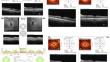
Results: OCT findings show postoperative changes in the eye: thinning in average and nasal circumpapillary retinal nerve fiber layer, thickening in macular central 1 mm inner plexus layer, ganglion cell complex, and nasal retinal nerve fiber layer. DTI reveals increased fractional anisotropy (FA) in the left optic chiasm and posterior optic nerve, decreased mid-segment optic nerve FA, and increased apparent diffusion coefficient (ADC) in the right optic chiasm and nerve segments. Early postoperative reduction in radial peripapillary capillaries plexus density is noted. Preoperative ganglion cell layer (GCL) thickness correlates with postoperative visual radiation FA and ADC values, especially in the inferior quadrant. A negative correlation exists between preoperative GCL thickness and postoperative visual field mean defect values, particularly on the temporal side and superior inner ring. All changes are statistically significant (P < 0.05).
Conclusions: The study finds that surgery for PA has varied effects on vision. Early post surgery, there are changes in the retina and nerve signals. Macular GCL thickness before surgery might predict early visual recovery, influencing future research and treatment for vision issues related to PA.
期刊介绍:
Aims and Scope
Ophthalmology and Therapy is an international, open access, peer-reviewed (single-blind), and rapid publication journal. The scope of the journal is broad and will consider all scientifically sound research from preclinical, clinical (all phases), observational, real-world, and health outcomes research around the use of ophthalmological therapies, devices, and surgical techniques.
The journal is of interest to a broad audience of pharmaceutical and healthcare professionals and publishes original research, reviews, case reports/series, trial protocols and short communications such as commentaries and editorials. Ophthalmology and Therapy will consider all scientifically sound research be it positive, confirmatory or negative data. Submissions are welcomed whether they relate to an international and/or a country-specific audience, something that is crucially important when researchers are trying to target more specific patient populations. This inclusive approach allows the journal to assist in the dissemination of quality research, which may be considered of insufficient interest by other journals.
Rapid Publication
The journal’s publication timelines aim for a rapid peer review of 2 weeks. If an article is accepted it will be published 3–4 weeks from acceptance. The rapid timelines are achieved through the combination of a dedicated in-house editorial team, who manage article workflow, and an extensive Editorial and Advisory Board who assist with peer review. This allows the journal to support the rapid dissemination of research, whilst still providing robust peer review. Combined with the journal’s open access model this allows for the rapid, efficient communication of the latest research and reviews, fostering the advancement of ophthalmic therapies.
Open Access
All articles published by Ophthalmology and Therapy are open access.
Personal Service
The journal’s dedicated in-house editorial team offer a personal “concierge service” meaning authors will always have an editorial contact able to update them on the status of their manuscript. The editorial team check all manuscripts to ensure that articles conform to the most recent COPE, GPP and ICMJE publishing guidelines. This supports the publication of ethically sound and transparent research.
Digital Features and Plain Language Summaries
Ophthalmology and Therapy offers a range of additional features designed to increase the visibility, readership and educational value of the journal’s content. Each article is accompanied by key summary points, giving a time-efficient overview of the content to a wide readership. Articles may be accompanied by plain language summaries to assist readers who have some knowledge of, but not in-depth expertise in, the area to understand the scientific content and overall implications of the article. The journal also provides the option to include various types of digital features including animated abstracts, video abstracts, slide decks, audio slides, instructional videos, infographics, podcasts and animations. All additional features are peer reviewed to the same high standard as the article itself. If you consider that your paper would benefit from the inclusion of a digital feature, please let us know. Our editorial team are able to create high-quality slide decks and infographics in-house, and video abstracts through our partner Research Square, and would be happy to assist in any way we can. For further information about digital features, please contact the journal editor (see ‘Contact the Journal’ for email address), and see the ‘Guidelines for digital features and plain language summaries’ document under ‘Submission guidelines’.
For examples of digital features please visit our showcase page https://springerhealthcare.com/expertise/publishing-digital-features/
Publication Fees
Upon acceptance of an article, authors will be required to pay the mandatory Rapid Service Fee of €5250/$6000/£4300. The journal will consider fee discounts and waivers for developing countries and this is decided on a case by case basis.
Peer Review Process
Upon submission, manuscripts are assessed by the editorial team to ensure they fit within the aims and scope of the journal and are also checked for plagiarism. All suitable submissions are then subject to a comprehensive single-blind peer review. Reviewers are selected based on their relevant expertise and publication history in the subject area. The journal has an extensive pool of editorial and advisory board members who have been selected to assist with peer review based on the afore-mentioned criteria.
At least two extensive reviews are required to make the editorial decision, with the exception of some article types such as Commentaries, Editorials, and Letters which are generally reviewed by one member of the Editorial Board. Where reviewer recommendations are conflicted, the editorial board will be contacted for further advice and a presiding decision. Manuscripts are then either accepted, rejected or authors are required to make major or minor revisions (both reviewer comments and editorial comments may need to be addressed). Once a revised manuscript is re-submitted, it is assessed along with the responses to reviewer comments and if it has been adequately revised it will be accepted for publication. Accepted manuscripts are then copyedited and typeset by the production team before online publication. Appeals against decisions following peer review are considered on a case-by-case basis and should be sent to the journal editor.
Preprints
We encourage posting of preprints of primary research manuscripts on preprint servers, authors’ or institutional websites, and open communications between researchers whether on community preprint servers or preprint commenting platforms. Posting of preprints is not considered prior publication and will not jeopardize consideration in our journals. Authors should disclose details of preprint posting during the submission process or at any other point during consideration in one of our journals. Once the manuscript is published, it is the author’s responsibility to ensure that the preprint record is updated with a publication reference, including the DOI and a URL link to the published version of the article on the journal website.
Please follow the link for further information on preprint sharing:
https://www.springer.com/gp/authors-editors/journal-author/journal-author-helpdesk/submission/1302#c16721550
Copyright
Ophthalmology and Therapy''s content is published open access under the Creative Commons Attribution-Noncommercial License, which allows users to read, copy, distribute, and make derivative works for non-commercial purposes from the material, as long as the author of the original work is cited. The author assigns the exclusive right to any commercial use of the article to Springer. For more information about the Creative Commons Attribution-Noncommercial License, click here: http://creativecommons.org/licenses/by-nc/4.0.
Contact
For more information about the journal, including pre-submission enquiries, please contact christopher.vautrinot@springer.com.

 求助内容:
求助内容: 应助结果提醒方式:


