{"title":"激活 P2Y2 受体可在肌肉再支配过程中促进神经肌肉接头的形成","authors":"Xianmin Song, Yingna Gao, Wei Wang, Hongliang Zheng, Minhui Zhu, Meng Li, Shicai Chen","doi":"10.1134/s1819712424020144","DOIUrl":null,"url":null,"abstract":"<h3 data-test=\"abstract-sub-heading\">Abstract</h3><p>Extracellular adenosine 5′-triphosphate (ATP), as neurotransmitter, is known to be an activity-dependent signaling molecule that regulates synaptic signaling. It is known to be co-released with acetylcholine from synaptic vesicle. The P2Y2 receptor (P2Y2R) is present in gastrocnemius muscles and co-localizes with post-synaptic acetylcholine receptors (AChRs). Accumulating evidence indicates that P2Y2R plays crucial roles in assembling neuromuscular junctions (NMJs), a peripheral synapse characterized by the clustering of AChRs on post-synaptic densities. This study investigated the alterations in P2Y2R expression during muscle reinnervation and the effect on NMJ formation. We found that P2Y2R consistently co-localized with AChR and sustained high expression in post-synaptic regions during muscle reinnervation. Notably, PSB1114-dependent stimulation of P2Y2R promoted NMJ formation and muscle reinnervation. The stimulatory effect of PSB1114 was significantly blocked by administration of the P2Y2R antagonist suramin. This study revealed distinctive patterns of expression and localization and a potential role of P2Y2R in promoting NMJ formation and regeneration during skeletal muscle reinnervation.</p>","PeriodicalId":19119,"journal":{"name":"Neurochemical Journal","volume":"58 1","pages":""},"PeriodicalIF":0.5000,"publicationDate":"2024-05-27","publicationTypes":"Journal Article","fieldsOfStudy":null,"isOpenAccess":false,"openAccessPdf":"","citationCount":"0","resultStr":"{\"title\":\"Activation of P2Y2 Receptors Promotes Neuromuscular Junction Formation during Muscle Reinnervation\",\"authors\":\"Xianmin Song, Yingna Gao, Wei Wang, Hongliang Zheng, Minhui Zhu, Meng Li, Shicai Chen\",\"doi\":\"10.1134/s1819712424020144\",\"DOIUrl\":null,\"url\":null,\"abstract\":\"<h3 data-test=\\\"abstract-sub-heading\\\">Abstract</h3><p>Extracellular adenosine 5′-triphosphate (ATP), as neurotransmitter, is known to be an activity-dependent signaling molecule that regulates synaptic signaling. It is known to be co-released with acetylcholine from synaptic vesicle. The P2Y2 receptor (P2Y2R) is present in gastrocnemius muscles and co-localizes with post-synaptic acetylcholine receptors (AChRs). Accumulating evidence indicates that P2Y2R plays crucial roles in assembling neuromuscular junctions (NMJs), a peripheral synapse characterized by the clustering of AChRs on post-synaptic densities. This study investigated the alterations in P2Y2R expression during muscle reinnervation and the effect on NMJ formation. We found that P2Y2R consistently co-localized with AChR and sustained high expression in post-synaptic regions during muscle reinnervation. Notably, PSB1114-dependent stimulation of P2Y2R promoted NMJ formation and muscle reinnervation. The stimulatory effect of PSB1114 was significantly blocked by administration of the P2Y2R antagonist suramin. This study revealed distinctive patterns of expression and localization and a potential role of P2Y2R in promoting NMJ formation and regeneration during skeletal muscle reinnervation.</p>\",\"PeriodicalId\":19119,\"journal\":{\"name\":\"Neurochemical Journal\",\"volume\":\"58 1\",\"pages\":\"\"},\"PeriodicalIF\":0.5000,\"publicationDate\":\"2024-05-27\",\"publicationTypes\":\"Journal Article\",\"fieldsOfStudy\":null,\"isOpenAccess\":false,\"openAccessPdf\":\"\",\"citationCount\":\"0\",\"resultStr\":null,\"platform\":\"Semanticscholar\",\"paperid\":null,\"PeriodicalName\":\"Neurochemical Journal\",\"FirstCategoryId\":\"3\",\"ListUrlMain\":\"https://doi.org/10.1134/s1819712424020144\",\"RegionNum\":4,\"RegionCategory\":\"医学\",\"ArticlePicture\":[],\"TitleCN\":null,\"AbstractTextCN\":null,\"PMCID\":null,\"EPubDate\":\"\",\"PubModel\":\"\",\"JCR\":\"Q4\",\"JCRName\":\"NEUROSCIENCES\",\"Score\":null,\"Total\":0}","platform":"Semanticscholar","paperid":null,"PeriodicalName":"Neurochemical Journal","FirstCategoryId":"3","ListUrlMain":"https://doi.org/10.1134/s1819712424020144","RegionNum":4,"RegionCategory":"医学","ArticlePicture":[],"TitleCN":null,"AbstractTextCN":null,"PMCID":null,"EPubDate":"","PubModel":"","JCR":"Q4","JCRName":"NEUROSCIENCES","Score":null,"Total":0}
Activation of P2Y2 Receptors Promotes Neuromuscular Junction Formation during Muscle Reinnervation
Abstract
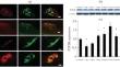
Extracellular adenosine 5′-triphosphate (ATP), as neurotransmitter, is known to be an activity-dependent signaling molecule that regulates synaptic signaling. It is known to be co-released with acetylcholine from synaptic vesicle. The P2Y2 receptor (P2Y2R) is present in gastrocnemius muscles and co-localizes with post-synaptic acetylcholine receptors (AChRs). Accumulating evidence indicates that P2Y2R plays crucial roles in assembling neuromuscular junctions (NMJs), a peripheral synapse characterized by the clustering of AChRs on post-synaptic densities. This study investigated the alterations in P2Y2R expression during muscle reinnervation and the effect on NMJ formation. We found that P2Y2R consistently co-localized with AChR and sustained high expression in post-synaptic regions during muscle reinnervation. Notably, PSB1114-dependent stimulation of P2Y2R promoted NMJ formation and muscle reinnervation. The stimulatory effect of PSB1114 was significantly blocked by administration of the P2Y2R antagonist suramin. This study revealed distinctive patterns of expression and localization and a potential role of P2Y2R in promoting NMJ formation and regeneration during skeletal muscle reinnervation.
期刊介绍:
Neurochemical Journal (Neirokhimiya) provides a source for the communication of the latest findings in all areas of contemporary neurochemistry and other fields of relevance (including molecular biology, biochemistry, physiology, neuroimmunology, pharmacology) in an afford to expand our understanding of the functions of the nervous system. The journal presents papers on functional neurochemistry, nervous system receptors, neurotransmitters, myelin, chromaffin granules and other components of the nervous system, as well as neurophysiological and clinical aspects, behavioral reactions, etc. Relevant topics include structure and function of the nervous system proteins, neuropeptides, nucleic acids, nucleotides, lipids, and other biologically active components.
The journal is devoted to the rapid publication of regular papers containing the results of original research, reviews highlighting major developments in neurochemistry, short communications, new experimental studies that use neurochemical methodology, descriptions of new methods of value for neurochemistry, theoretical material suggesting novel principles and approaches to neurochemical problems, presentations of new hypotheses and significant findings, discussions, chronicles of congresses, meetings, and conferences with short presentations of the most sensational and timely reports, information on the activity of the Russian and International Neurochemical Societies, as well as advertisements of reagents and equipment.
 求助内容:
求助内容: 应助结果提醒方式:


