Patrick H Luckett, Michael O Olufawo, Ki Yun Park, Bidhan Lamichhane, Donna Dierker, Gabriel Trevino Verastegui, John J Lee, Peter Yang, Albert Kim, Omar H Butt, Milan G Chheda, Abraham Z Snyder, Joshua S Shimony, Eric C Leuthardt
{"title":"利用静息状态 fMRI 和机器学习预测高级别胶质瘤手术后的功能状态。","authors":"Patrick H Luckett, Michael O Olufawo, Ki Yun Park, Bidhan Lamichhane, Donna Dierker, Gabriel Trevino Verastegui, John J Lee, Peter Yang, Albert Kim, Omar H Butt, Milan G Chheda, Abraham Z Snyder, Joshua S Shimony, Eric C Leuthardt","doi":"10.1007/s11060-024-04715-1","DOIUrl":null,"url":null,"abstract":"<p><strong>Purpose: </strong>High-grade glioma (HGG) is the most common and deadly malignant glioma of the central nervous system. The current standard of care includes surgical resection of the tumor, which can lead to functional and cognitive deficits. The aim of this study is to develop models capable of predicting functional outcomes in HGG patients before surgery, facilitating improved disease management and informed patient care.</p><p><strong>Methods: </strong>Adult HGG patients (N = 102) from the neurosurgery brain tumor service at Washington University Medical Center were retrospectively recruited. All patients completed structural neuroimaging and resting state functional MRI prior to surgery. Demographics, measures of resting state network connectivity (FC), tumor location, and tumor volume were used to train a random forest classifier to predict functional outcomes based on Karnofsky Performance Status (KPS < 70, KPS ≥ 70).</p><p><strong>Results: </strong>The models achieved a nested cross-validation accuracy of 94.1% and an AUC of 0.97 in classifying KPS. The strongest predictors identified by the model included FC between somatomotor, visual, auditory, and reward networks. Based on location, the relation of the tumor to dorsal attention, cingulo-opercular, and basal ganglia networks were strong predictors of KPS. Age was also a strong predictor. However, tumor volume was only a moderate predictor.</p><p><strong>Conclusion: </strong>The current work demonstrates the ability of machine learning to classify postoperative functional outcomes in HGG patients prior to surgery accurately. Our results suggest that both FC and the tumor's location in relation to specific networks can serve as reliable predictors of functional outcomes, leading to personalized therapeutic approaches tailored to individual patients.</p>","PeriodicalId":16425,"journal":{"name":"Journal of Neuro-Oncology","volume":" ","pages":"175-185"},"PeriodicalIF":3.2000,"publicationDate":"2024-08-01","publicationTypes":"Journal Article","fieldsOfStudy":null,"isOpenAccess":false,"openAccessPdf":"https://www.ncbi.nlm.nih.gov/pmc/articles/PMC11269343/pdf/","citationCount":"0","resultStr":"{\"title\":\"Predicting post-surgical functional status in high-grade glioma with resting state fMRI and machine learning.\",\"authors\":\"Patrick H Luckett, Michael O Olufawo, Ki Yun Park, Bidhan Lamichhane, Donna Dierker, Gabriel Trevino Verastegui, John J Lee, Peter Yang, Albert Kim, Omar H Butt, Milan G Chheda, Abraham Z Snyder, Joshua S Shimony, Eric C Leuthardt\",\"doi\":\"10.1007/s11060-024-04715-1\",\"DOIUrl\":null,\"url\":null,\"abstract\":\"<p><strong>Purpose: </strong>High-grade glioma (HGG) is the most common and deadly malignant glioma of the central nervous system. The current standard of care includes surgical resection of the tumor, which can lead to functional and cognitive deficits. The aim of this study is to develop models capable of predicting functional outcomes in HGG patients before surgery, facilitating improved disease management and informed patient care.</p><p><strong>Methods: </strong>Adult HGG patients (N = 102) from the neurosurgery brain tumor service at Washington University Medical Center were retrospectively recruited. All patients completed structural neuroimaging and resting state functional MRI prior to surgery. Demographics, measures of resting state network connectivity (FC), tumor location, and tumor volume were used to train a random forest classifier to predict functional outcomes based on Karnofsky Performance Status (KPS < 70, KPS ≥ 70).</p><p><strong>Results: </strong>The models achieved a nested cross-validation accuracy of 94.1% and an AUC of 0.97 in classifying KPS. The strongest predictors identified by the model included FC between somatomotor, visual, auditory, and reward networks. Based on location, the relation of the tumor to dorsal attention, cingulo-opercular, and basal ganglia networks were strong predictors of KPS. Age was also a strong predictor. However, tumor volume was only a moderate predictor.</p><p><strong>Conclusion: </strong>The current work demonstrates the ability of machine learning to classify postoperative functional outcomes in HGG patients prior to surgery accurately. Our results suggest that both FC and the tumor's location in relation to specific networks can serve as reliable predictors of functional outcomes, leading to personalized therapeutic approaches tailored to individual patients.</p>\",\"PeriodicalId\":16425,\"journal\":{\"name\":\"Journal of Neuro-Oncology\",\"volume\":\" \",\"pages\":\"175-185\"},\"PeriodicalIF\":3.2000,\"publicationDate\":\"2024-08-01\",\"publicationTypes\":\"Journal Article\",\"fieldsOfStudy\":null,\"isOpenAccess\":false,\"openAccessPdf\":\"https://www.ncbi.nlm.nih.gov/pmc/articles/PMC11269343/pdf/\",\"citationCount\":\"0\",\"resultStr\":null,\"platform\":\"Semanticscholar\",\"paperid\":null,\"PeriodicalName\":\"Journal of Neuro-Oncology\",\"FirstCategoryId\":\"3\",\"ListUrlMain\":\"https://doi.org/10.1007/s11060-024-04715-1\",\"RegionNum\":2,\"RegionCategory\":\"医学\",\"ArticlePicture\":[],\"TitleCN\":null,\"AbstractTextCN\":null,\"PMCID\":null,\"EPubDate\":\"2024/5/24 0:00:00\",\"PubModel\":\"Epub\",\"JCR\":\"Q2\",\"JCRName\":\"CLINICAL NEUROLOGY\",\"Score\":null,\"Total\":0}","platform":"Semanticscholar","paperid":null,"PeriodicalName":"Journal of Neuro-Oncology","FirstCategoryId":"3","ListUrlMain":"https://doi.org/10.1007/s11060-024-04715-1","RegionNum":2,"RegionCategory":"医学","ArticlePicture":[],"TitleCN":null,"AbstractTextCN":null,"PMCID":null,"EPubDate":"2024/5/24 0:00:00","PubModel":"Epub","JCR":"Q2","JCRName":"CLINICAL NEUROLOGY","Score":null,"Total":0}
Predicting post-surgical functional status in high-grade glioma with resting state fMRI and machine learning.
Purpose: High-grade glioma (HGG) is the most common and deadly malignant glioma of the central nervous system. The current standard of care includes surgical resection of the tumor, which can lead to functional and cognitive deficits. The aim of this study is to develop models capable of predicting functional outcomes in HGG patients before surgery, facilitating improved disease management and informed patient care.
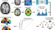
Methods: Adult HGG patients (N = 102) from the neurosurgery brain tumor service at Washington University Medical Center were retrospectively recruited. All patients completed structural neuroimaging and resting state functional MRI prior to surgery. Demographics, measures of resting state network connectivity (FC), tumor location, and tumor volume were used to train a random forest classifier to predict functional outcomes based on Karnofsky Performance Status (KPS < 70, KPS ≥ 70).
Results: The models achieved a nested cross-validation accuracy of 94.1% and an AUC of 0.97 in classifying KPS. The strongest predictors identified by the model included FC between somatomotor, visual, auditory, and reward networks. Based on location, the relation of the tumor to dorsal attention, cingulo-opercular, and basal ganglia networks were strong predictors of KPS. Age was also a strong predictor. However, tumor volume was only a moderate predictor.
Conclusion: The current work demonstrates the ability of machine learning to classify postoperative functional outcomes in HGG patients prior to surgery accurately. Our results suggest that both FC and the tumor's location in relation to specific networks can serve as reliable predictors of functional outcomes, leading to personalized therapeutic approaches tailored to individual patients.
期刊介绍:
The Journal of Neuro-Oncology is a multi-disciplinary journal encompassing basic, applied, and clinical investigations in all research areas as they relate to cancer and the central nervous system. It provides a single forum for communication among neurologists, neurosurgeons, radiotherapists, medical oncologists, neuropathologists, neurodiagnosticians, and laboratory-based oncologists conducting relevant research. The Journal of Neuro-Oncology does not seek to isolate the field, but rather to focus the efforts of many disciplines in one publication through a format which pulls together these diverse interests. More than any other field of oncology, cancer of the central nervous system requires multi-disciplinary approaches. To alleviate having to scan dozens of journals of cell biology, pathology, laboratory and clinical endeavours, JNO is a periodical in which current, high-quality, relevant research in all aspects of neuro-oncology may be found.

 求助内容:
求助内容: 应助结果提醒方式:


