Jeffrey Strakowski, Han Zhang, Millard Reschke, Faye Y Chiou-Tan
{"title":"特殊解剖系列。利用高频超声波成像内耳结构:应用于物理康复空间医学。","authors":"Jeffrey Strakowski, Han Zhang, Millard Reschke, Faye Y Chiou-Tan","doi":"10.1097/ph9.0000000000000026","DOIUrl":null,"url":null,"abstract":"<p><strong>Objective: </strong>The objective of this paper is to document the feasibility of image acquisition, image optimization, and sonographic appearance of the exposed anatomic windows of cadaveric inner ear dissection for purposes of potential future clinical evaluation as part of the developing area of physical and rehabilitation space medicine.</p><p><strong>Methods: </strong>Cadaveric dissection of the inner ear was conducted with the goal of exposing areas relevant to vestibular balance. Middle and inner ear structures of 3 human cadavers were imaged with multiple broadband transducers, including emphasis with higher frequency transducers.</p><p><strong>Results: </strong>The images were best optimized with 17 MHz and 22 MHz small footprint transducers. High-frequency ultrasound (US) images of the semicircular canals, vestibular and facial nerves, and utricles with reflected otoliths (otoconia) were obtained and reported in this article. Detailed visualization of both the vestibular nerve and facial nerve was accomplished, including identification of fascicular architecture. In addition, US reflection from the otoliths contained within the utricle was identified with sufficient clarity to provide surface measurements. Bony acoustic landmarks of the middle ear bones were identified by scanning externally from the tympanic membrane, including the dynamic movement of the bones with manual manipulation.</p><p><strong>Conclusion: </strong>US visualization has the potential to be an effective imaging modality to monitor potential changes to the otolith's size throughout extended space flight. To our knowledge, no prior study has reported US images of human inner ear structures.</p>","PeriodicalId":75125,"journal":{"name":"The journal of the International Society of Physical and Rehabilitation Medicine","volume":"7 1","pages":"33-38"},"PeriodicalIF":0.0000,"publicationDate":"2024-01-25","publicationTypes":"Journal Article","fieldsOfStudy":null,"isOpenAccess":false,"openAccessPdf":"https://www.ncbi.nlm.nih.gov/pmc/articles/PMC11111317/pdf/","citationCount":"0","resultStr":"{\"title\":\"Special anatomy series. Imaging inner ear structures with high-frequency ultrasound: Application to physical rehabilitation space medicine.\",\"authors\":\"Jeffrey Strakowski, Han Zhang, Millard Reschke, Faye Y Chiou-Tan\",\"doi\":\"10.1097/ph9.0000000000000026\",\"DOIUrl\":null,\"url\":null,\"abstract\":\"<p><strong>Objective: </strong>The objective of this paper is to document the feasibility of image acquisition, image optimization, and sonographic appearance of the exposed anatomic windows of cadaveric inner ear dissection for purposes of potential future clinical evaluation as part of the developing area of physical and rehabilitation space medicine.</p><p><strong>Methods: </strong>Cadaveric dissection of the inner ear was conducted with the goal of exposing areas relevant to vestibular balance. Middle and inner ear structures of 3 human cadavers were imaged with multiple broadband transducers, including emphasis with higher frequency transducers.</p><p><strong>Results: </strong>The images were best optimized with 17 MHz and 22 MHz small footprint transducers. High-frequency ultrasound (US) images of the semicircular canals, vestibular and facial nerves, and utricles with reflected otoliths (otoconia) were obtained and reported in this article. Detailed visualization of both the vestibular nerve and facial nerve was accomplished, including identification of fascicular architecture. In addition, US reflection from the otoliths contained within the utricle was identified with sufficient clarity to provide surface measurements. Bony acoustic landmarks of the middle ear bones were identified by scanning externally from the tympanic membrane, including the dynamic movement of the bones with manual manipulation.</p><p><strong>Conclusion: </strong>US visualization has the potential to be an effective imaging modality to monitor potential changes to the otolith's size throughout extended space flight. To our knowledge, no prior study has reported US images of human inner ear structures.</p>\",\"PeriodicalId\":75125,\"journal\":{\"name\":\"The journal of the International Society of Physical and Rehabilitation Medicine\",\"volume\":\"7 1\",\"pages\":\"33-38\"},\"PeriodicalIF\":0.0000,\"publicationDate\":\"2024-01-25\",\"publicationTypes\":\"Journal Article\",\"fieldsOfStudy\":null,\"isOpenAccess\":false,\"openAccessPdf\":\"https://www.ncbi.nlm.nih.gov/pmc/articles/PMC11111317/pdf/\",\"citationCount\":\"0\",\"resultStr\":null,\"platform\":\"Semanticscholar\",\"paperid\":null,\"PeriodicalName\":\"The journal of the International Society of Physical and Rehabilitation Medicine\",\"FirstCategoryId\":\"1085\",\"ListUrlMain\":\"https://doi.org/10.1097/ph9.0000000000000026\",\"RegionNum\":0,\"RegionCategory\":null,\"ArticlePicture\":[],\"TitleCN\":null,\"AbstractTextCN\":null,\"PMCID\":null,\"EPubDate\":\"2024/5/1 0:00:00\",\"PubModel\":\"eCollection\",\"JCR\":\"\",\"JCRName\":\"\",\"Score\":null,\"Total\":0}","platform":"Semanticscholar","paperid":null,"PeriodicalName":"The journal of the International Society of Physical and Rehabilitation Medicine","FirstCategoryId":"1085","ListUrlMain":"https://doi.org/10.1097/ph9.0000000000000026","RegionNum":0,"RegionCategory":null,"ArticlePicture":[],"TitleCN":null,"AbstractTextCN":null,"PMCID":null,"EPubDate":"2024/5/1 0:00:00","PubModel":"eCollection","JCR":"","JCRName":"","Score":null,"Total":0}
Special anatomy series. Imaging inner ear structures with high-frequency ultrasound: Application to physical rehabilitation space medicine.
Objective: The objective of this paper is to document the feasibility of image acquisition, image optimization, and sonographic appearance of the exposed anatomic windows of cadaveric inner ear dissection for purposes of potential future clinical evaluation as part of the developing area of physical and rehabilitation space medicine.
Methods: Cadaveric dissection of the inner ear was conducted with the goal of exposing areas relevant to vestibular balance. Middle and inner ear structures of 3 human cadavers were imaged with multiple broadband transducers, including emphasis with higher frequency transducers.
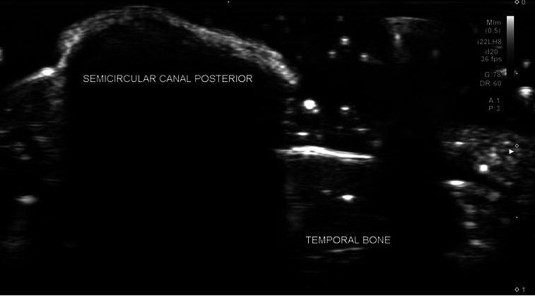
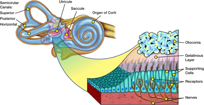
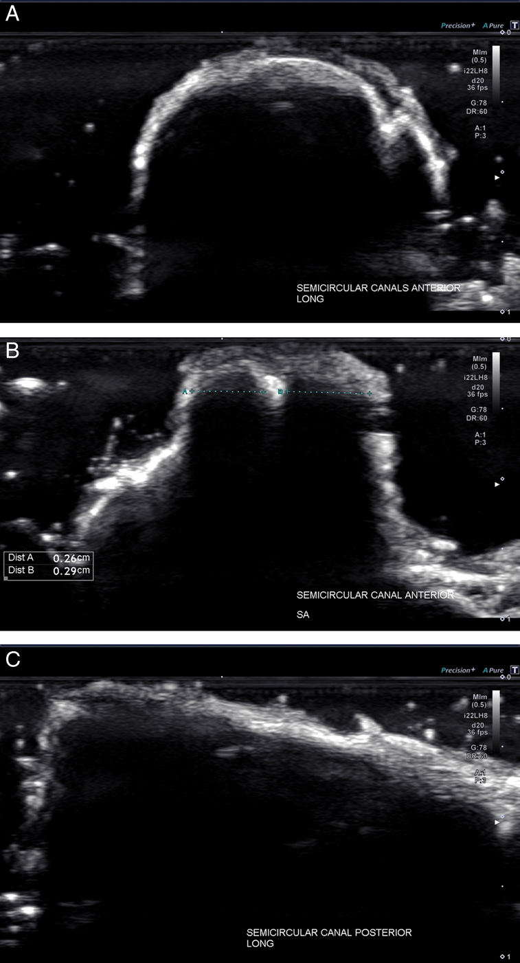
Results: The images were best optimized with 17 MHz and 22 MHz small footprint transducers. High-frequency ultrasound (US) images of the semicircular canals, vestibular and facial nerves, and utricles with reflected otoliths (otoconia) were obtained and reported in this article. Detailed visualization of both the vestibular nerve and facial nerve was accomplished, including identification of fascicular architecture. In addition, US reflection from the otoliths contained within the utricle was identified with sufficient clarity to provide surface measurements. Bony acoustic landmarks of the middle ear bones were identified by scanning externally from the tympanic membrane, including the dynamic movement of the bones with manual manipulation.
Conclusion: US visualization has the potential to be an effective imaging modality to monitor potential changes to the otolith's size throughout extended space flight. To our knowledge, no prior study has reported US images of human inner ear structures.

 求助内容:
求助内容: 应助结果提醒方式:


