Haoye Meng , Xuejian Liu , Ronghui Liu , Yudong Zheng , Angyang Hou , Shuyun Liu , Wei He , Yu Wang , Aiyuan Wang , Quanyi Guo , Jiang Peng
{"title":"脱细胞激光微图案骨软骨植入物在山羊模型中表现出分区再细胞化和自固定功能,促进骨软骨再生","authors":"Haoye Meng , Xuejian Liu , Ronghui Liu , Yudong Zheng , Angyang Hou , Shuyun Liu , Wei He , Yu Wang , Aiyuan Wang , Quanyi Guo , Jiang Peng","doi":"10.1016/j.jot.2024.04.005","DOIUrl":null,"url":null,"abstract":"<div><h3>Background</h3><p>Osteochondral regeneration has long been recognized as a complex and challenging project in the field of tissue engineering. In particular, reconstructing the osteochondral interface is crucial for determining the effectiveness of the repair. Although several artificial layered or gradient scaffolds have been developed recently to simulate the natural interface, the functions of this unique structure have still not been fully replicated. In this paper, we utilized laser micro-patterning technology (LMPT) to modify the natural osteochondral “plugs” for use as grafts and aimed to directly apply the functional interface unit to repair osteochondral defects in a goat model.</p></div><div><h3>Methods</h3><p>For in vitro evaluations, the optimal combination of LMPT parameters was confirmed through mechanical testing, finite element analysis, and comparing decellularization efficiency. The structural and biological properties of the laser micro-patterned osteochondral implants (LMP-OI) were verified by measuring the permeability of the interface and assessing the recellularization processes. In the goat model for osteochondral regeneration, a conical frustum-shaped defect was specifically created in the weight-bearing area of femoral condyles using a customized trephine with a variable diameter. This unreported defect shape enabled the implant to properly self-fix as expected.</p></div><div><h3>Results</h3><p>The micro-patterning with the suitable pore density and morphology increased the permeability of the LMP-OIs, accelerated decellularization, maintained mechanical stability, and provided two relative independent microenvironments for subsequent recellularization. The LMP-OIs with goat's autologous bone marrow stromal cells in the cartilage layer have securely integrated into the osteochondral defects. At 6 and 12 months after implantation, both imaging and histological assessments showed a significant improvement in the healing of the cartilage and subchondral bone.</p></div><div><h3>Conclusion</h3><p>With the natural interface unit and zonal recellularization, the LMP-OI is an ideal scaffold to repair osteochondral defects especially in large animals.</p></div><div><h3>The translational potential of this article</h3><p>These findings suggest that such a modified xenogeneic osteochondral implant could potentially be explored in clinical translation for treatment of osteochondral injuries. Furthermore, trimming a conical frustum shape to the defect region, especially for large-sized defects, may be an effective way to achieve self-fixing for the implant.</p></div>","PeriodicalId":16636,"journal":{"name":"Journal of Orthopaedic Translation","volume":"46 ","pages":"Pages 18-32"},"PeriodicalIF":5.9000,"publicationDate":"2024-05-01","publicationTypes":"Journal Article","fieldsOfStudy":null,"isOpenAccess":false,"openAccessPdf":"https://www.sciencedirect.com/science/article/pii/S2214031X24000421/pdfft?md5=accc0f1e9f084a3ffec7c5b50e01064e&pid=1-s2.0-S2214031X24000421-main.pdf","citationCount":"0","resultStr":"{\"title\":\"Decellularized laser micro-patterned osteochondral implants exhibit zonal recellularization and self-fixing for osteochondral regeneration in a goat model\",\"authors\":\"Haoye Meng , Xuejian Liu , Ronghui Liu , Yudong Zheng , Angyang Hou , Shuyun Liu , Wei He , Yu Wang , Aiyuan Wang , Quanyi Guo , Jiang Peng\",\"doi\":\"10.1016/j.jot.2024.04.005\",\"DOIUrl\":null,\"url\":null,\"abstract\":\"<div><h3>Background</h3><p>Osteochondral regeneration has long been recognized as a complex and challenging project in the field of tissue engineering. In particular, reconstructing the osteochondral interface is crucial for determining the effectiveness of the repair. Although several artificial layered or gradient scaffolds have been developed recently to simulate the natural interface, the functions of this unique structure have still not been fully replicated. In this paper, we utilized laser micro-patterning technology (LMPT) to modify the natural osteochondral “plugs” for use as grafts and aimed to directly apply the functional interface unit to repair osteochondral defects in a goat model.</p></div><div><h3>Methods</h3><p>For in vitro evaluations, the optimal combination of LMPT parameters was confirmed through mechanical testing, finite element analysis, and comparing decellularization efficiency. The structural and biological properties of the laser micro-patterned osteochondral implants (LMP-OI) were verified by measuring the permeability of the interface and assessing the recellularization processes. In the goat model for osteochondral regeneration, a conical frustum-shaped defect was specifically created in the weight-bearing area of femoral condyles using a customized trephine with a variable diameter. This unreported defect shape enabled the implant to properly self-fix as expected.</p></div><div><h3>Results</h3><p>The micro-patterning with the suitable pore density and morphology increased the permeability of the LMP-OIs, accelerated decellularization, maintained mechanical stability, and provided two relative independent microenvironments for subsequent recellularization. The LMP-OIs with goat's autologous bone marrow stromal cells in the cartilage layer have securely integrated into the osteochondral defects. At 6 and 12 months after implantation, both imaging and histological assessments showed a significant improvement in the healing of the cartilage and subchondral bone.</p></div><div><h3>Conclusion</h3><p>With the natural interface unit and zonal recellularization, the LMP-OI is an ideal scaffold to repair osteochondral defects especially in large animals.</p></div><div><h3>The translational potential of this article</h3><p>These findings suggest that such a modified xenogeneic osteochondral implant could potentially be explored in clinical translation for treatment of osteochondral injuries. Furthermore, trimming a conical frustum shape to the defect region, especially for large-sized defects, may be an effective way to achieve self-fixing for the implant.</p></div>\",\"PeriodicalId\":16636,\"journal\":{\"name\":\"Journal of Orthopaedic Translation\",\"volume\":\"46 \",\"pages\":\"Pages 18-32\"},\"PeriodicalIF\":5.9000,\"publicationDate\":\"2024-05-01\",\"publicationTypes\":\"Journal Article\",\"fieldsOfStudy\":null,\"isOpenAccess\":false,\"openAccessPdf\":\"https://www.sciencedirect.com/science/article/pii/S2214031X24000421/pdfft?md5=accc0f1e9f084a3ffec7c5b50e01064e&pid=1-s2.0-S2214031X24000421-main.pdf\",\"citationCount\":\"0\",\"resultStr\":null,\"platform\":\"Semanticscholar\",\"paperid\":null,\"PeriodicalName\":\"Journal of Orthopaedic Translation\",\"FirstCategoryId\":\"3\",\"ListUrlMain\":\"https://www.sciencedirect.com/science/article/pii/S2214031X24000421\",\"RegionNum\":1,\"RegionCategory\":\"医学\",\"ArticlePicture\":[],\"TitleCN\":null,\"AbstractTextCN\":null,\"PMCID\":null,\"EPubDate\":\"\",\"PubModel\":\"\",\"JCR\":\"Q1\",\"JCRName\":\"ORTHOPEDICS\",\"Score\":null,\"Total\":0}","platform":"Semanticscholar","paperid":null,"PeriodicalName":"Journal of Orthopaedic Translation","FirstCategoryId":"3","ListUrlMain":"https://www.sciencedirect.com/science/article/pii/S2214031X24000421","RegionNum":1,"RegionCategory":"医学","ArticlePicture":[],"TitleCN":null,"AbstractTextCN":null,"PMCID":null,"EPubDate":"","PubModel":"","JCR":"Q1","JCRName":"ORTHOPEDICS","Score":null,"Total":0}
Decellularized laser micro-patterned osteochondral implants exhibit zonal recellularization and self-fixing for osteochondral regeneration in a goat model
Background
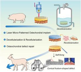
Osteochondral regeneration has long been recognized as a complex and challenging project in the field of tissue engineering. In particular, reconstructing the osteochondral interface is crucial for determining the effectiveness of the repair. Although several artificial layered or gradient scaffolds have been developed recently to simulate the natural interface, the functions of this unique structure have still not been fully replicated. In this paper, we utilized laser micro-patterning technology (LMPT) to modify the natural osteochondral “plugs” for use as grafts and aimed to directly apply the functional interface unit to repair osteochondral defects in a goat model.
Methods
For in vitro evaluations, the optimal combination of LMPT parameters was confirmed through mechanical testing, finite element analysis, and comparing decellularization efficiency. The structural and biological properties of the laser micro-patterned osteochondral implants (LMP-OI) were verified by measuring the permeability of the interface and assessing the recellularization processes. In the goat model for osteochondral regeneration, a conical frustum-shaped defect was specifically created in the weight-bearing area of femoral condyles using a customized trephine with a variable diameter. This unreported defect shape enabled the implant to properly self-fix as expected.
Results
The micro-patterning with the suitable pore density and morphology increased the permeability of the LMP-OIs, accelerated decellularization, maintained mechanical stability, and provided two relative independent microenvironments for subsequent recellularization. The LMP-OIs with goat's autologous bone marrow stromal cells in the cartilage layer have securely integrated into the osteochondral defects. At 6 and 12 months after implantation, both imaging and histological assessments showed a significant improvement in the healing of the cartilage and subchondral bone.
Conclusion
With the natural interface unit and zonal recellularization, the LMP-OI is an ideal scaffold to repair osteochondral defects especially in large animals.
The translational potential of this article
These findings suggest that such a modified xenogeneic osteochondral implant could potentially be explored in clinical translation for treatment of osteochondral injuries. Furthermore, trimming a conical frustum shape to the defect region, especially for large-sized defects, may be an effective way to achieve self-fixing for the implant.
期刊介绍:
The Journal of Orthopaedic Translation (JOT) is the official peer-reviewed, open access journal of the Chinese Speaking Orthopaedic Society (CSOS) and the International Chinese Musculoskeletal Research Society (ICMRS). It is published quarterly, in January, April, July and October, by Elsevier.
 求助内容:
求助内容: 应助结果提醒方式:


