{"title":"[骨髓间充质干细胞提取的外泌体通过 miR-335 调控 NF-κB 通路并减轻大鼠肺缺血再灌注损伤]","authors":"Bing Zhang, Chao Meng, Ji-Yu Kang, Hua-Cheng Zhou","doi":"","DOIUrl":null,"url":null,"abstract":"<p><p>This study aimed to investigate the effect of exosomes derived from bone marrow mesenchymal stem cells (BMSCs-EXO) on lung ischemia-reperfusion injury (IRI) in rats and to explore the role of miR-335. The model of rat lung IRI was established by clipping the hilum of left lung for 60 min and opening for 180 min. Forty Sprague-Dawley rats were randomly divided into sham group, IRI group, IRI+PBS group, IRI+EXO group, and IRI+miR-335 inhibitor EXO (IRI+inhibitor-EXO) group (n = 8). Rats in the sham group underwent thoracotomies without IRI. Rats in the IRI group were used to establish IRI model without any additional treatment. In the IRI+PBS, IRI+EXO, and IRI+inhibitor-EXO groups, the rats were used to establish IRI model and given PBS, EXO from BMSCs without any treatment, and EXO from BMSCs with miR-335 inhibitor treatment before reperfusion, respectively. Blood gases were analyzed during the experiment. Lung tissue wet/dry ratio (W/D), interleukin 1β (IL-1β), tumor necrosis factor α (TNF-α), myeloperoxidase (MPO), malondialdehyde (MDA), and superoxide dismutase (SOD) were measured at the end of reperfusion. Mitochondria were observed by electron microscopy and the Flameng scores were counted. Lung histopathology and apoptosis (TUNEL staining) were observed by light microscopy, and the lung injury scores (LIS) and apoptosis index (AI) were detected. The miR-335 expression was detected by RT-qPCR, and the expression of caspase-3, cleaved-caspase-3, caspase-9, cleaved-caspase-9, and NF-κB proteins were detected by Western blot at the end of reperfusion. The results showed that compared with the sham group, the oxygenation index, pH, and base excess (BE) were significantly lower in the IRI group and IRI+PBS group after reperfusion, whereas those indices were significantly higher in the IRI+EXO group than those in the IRI+PBS group (P < 0.05). Compared with the sham group, there were significant increases in W/D, IL-1β, TNF-α, MPO, MDA, LIS, AI, Flameng score, caspase-3, cleaved-caspase-3, caspase-9, and cleaved-caspase-9, however significant decreases in the SOD, miR-335 and NF-κB in the IRI group (P < 0.05). These indices in the IRI and IRI+PBS groups showed no significant differences. Compared with the IRI+PBS group, there were significant decreases in W/D, IL-1β, TNF-α, MPO, MDA, LIS, AI, Flameng score, caspase-3, cleaved-caspase-3, caspase-9, and cleaved-caspase-9, however significant increases in the SOD, miR-335 and NF-κB in the IRI+EXO group (P < 0.05). While, the changes of the above mentioned indices were reversed in the IRI+inhibitor-EXO group compared with IRI+EXO group, which were still better than those in the IRI+PBS group (P < 0.05). The results suggest that BMSCs-EXO could attenuate lung IRI in rats, activate NF-κB pathway, and maintain mitochondrial stability by up-regulating miR-335.</p>","PeriodicalId":7134,"journal":{"name":"生理学报","volume":null,"pages":null},"PeriodicalIF":0.0000,"publicationDate":"2024-04-25","publicationTypes":"Journal Article","fieldsOfStudy":null,"isOpenAccess":false,"openAccessPdf":"","citationCount":"0","resultStr":"{\"title\":\"[Exosomes derived from bone marrow mesenchymal stem cells regulate NF-κB pathway and reduce lung ischemia-reperfusion injury in rats by miR-335].\",\"authors\":\"Bing Zhang, Chao Meng, Ji-Yu Kang, Hua-Cheng Zhou\",\"doi\":\"\",\"DOIUrl\":null,\"url\":null,\"abstract\":\"<p><p>This study aimed to investigate the effect of exosomes derived from bone marrow mesenchymal stem cells (BMSCs-EXO) on lung ischemia-reperfusion injury (IRI) in rats and to explore the role of miR-335. The model of rat lung IRI was established by clipping the hilum of left lung for 60 min and opening for 180 min. Forty Sprague-Dawley rats were randomly divided into sham group, IRI group, IRI+PBS group, IRI+EXO group, and IRI+miR-335 inhibitor EXO (IRI+inhibitor-EXO) group (n = 8). Rats in the sham group underwent thoracotomies without IRI. Rats in the IRI group were used to establish IRI model without any additional treatment. In the IRI+PBS, IRI+EXO, and IRI+inhibitor-EXO groups, the rats were used to establish IRI model and given PBS, EXO from BMSCs without any treatment, and EXO from BMSCs with miR-335 inhibitor treatment before reperfusion, respectively. Blood gases were analyzed during the experiment. Lung tissue wet/dry ratio (W/D), interleukin 1β (IL-1β), tumor necrosis factor α (TNF-α), myeloperoxidase (MPO), malondialdehyde (MDA), and superoxide dismutase (SOD) were measured at the end of reperfusion. Mitochondria were observed by electron microscopy and the Flameng scores were counted. Lung histopathology and apoptosis (TUNEL staining) were observed by light microscopy, and the lung injury scores (LIS) and apoptosis index (AI) were detected. The miR-335 expression was detected by RT-qPCR, and the expression of caspase-3, cleaved-caspase-3, caspase-9, cleaved-caspase-9, and NF-κB proteins were detected by Western blot at the end of reperfusion. The results showed that compared with the sham group, the oxygenation index, pH, and base excess (BE) were significantly lower in the IRI group and IRI+PBS group after reperfusion, whereas those indices were significantly higher in the IRI+EXO group than those in the IRI+PBS group (P < 0.05). Compared with the sham group, there were significant increases in W/D, IL-1β, TNF-α, MPO, MDA, LIS, AI, Flameng score, caspase-3, cleaved-caspase-3, caspase-9, and cleaved-caspase-9, however significant decreases in the SOD, miR-335 and NF-κB in the IRI group (P < 0.05). These indices in the IRI and IRI+PBS groups showed no significant differences. Compared with the IRI+PBS group, there were significant decreases in W/D, IL-1β, TNF-α, MPO, MDA, LIS, AI, Flameng score, caspase-3, cleaved-caspase-3, caspase-9, and cleaved-caspase-9, however significant increases in the SOD, miR-335 and NF-κB in the IRI+EXO group (P < 0.05). While, the changes of the above mentioned indices were reversed in the IRI+inhibitor-EXO group compared with IRI+EXO group, which were still better than those in the IRI+PBS group (P < 0.05). The results suggest that BMSCs-EXO could attenuate lung IRI in rats, activate NF-κB pathway, and maintain mitochondrial stability by up-regulating miR-335.</p>\",\"PeriodicalId\":7134,\"journal\":{\"name\":\"生理学报\",\"volume\":null,\"pages\":null},\"PeriodicalIF\":0.0000,\"publicationDate\":\"2024-04-25\",\"publicationTypes\":\"Journal Article\",\"fieldsOfStudy\":null,\"isOpenAccess\":false,\"openAccessPdf\":\"\",\"citationCount\":\"0\",\"resultStr\":null,\"platform\":\"Semanticscholar\",\"paperid\":null,\"PeriodicalName\":\"生理学报\",\"FirstCategoryId\":\"1087\",\"ListUrlMain\":\"\",\"RegionNum\":0,\"RegionCategory\":null,\"ArticlePicture\":[],\"TitleCN\":null,\"AbstractTextCN\":null,\"PMCID\":null,\"EPubDate\":\"\",\"PubModel\":\"\",\"JCR\":\"Q3\",\"JCRName\":\"Medicine\",\"Score\":null,\"Total\":0}","platform":"Semanticscholar","paperid":null,"PeriodicalName":"生理学报","FirstCategoryId":"1087","ListUrlMain":"","RegionNum":0,"RegionCategory":null,"ArticlePicture":[],"TitleCN":null,"AbstractTextCN":null,"PMCID":null,"EPubDate":"","PubModel":"","JCR":"Q3","JCRName":"Medicine","Score":null,"Total":0}
[Exosomes derived from bone marrow mesenchymal stem cells regulate NF-κB pathway and reduce lung ischemia-reperfusion injury in rats by miR-335].
This study aimed to investigate the effect of exosomes derived from bone marrow mesenchymal stem cells (BMSCs-EXO) on lung ischemia-reperfusion injury (IRI) in rats and to explore the role of miR-335. The model of rat lung IRI was established by clipping the hilum of left lung for 60 min and opening for 180 min. Forty Sprague-Dawley rats were randomly divided into sham group, IRI group, IRI+PBS group, IRI+EXO group, and IRI+miR-335 inhibitor EXO (IRI+inhibitor-EXO) group (n = 8). Rats in the sham group underwent thoracotomies without IRI. Rats in the IRI group were used to establish IRI model without any additional treatment. In the IRI+PBS, IRI+EXO, and IRI+inhibitor-EXO groups, the rats were used to establish IRI model and given PBS, EXO from BMSCs without any treatment, and EXO from BMSCs with miR-335 inhibitor treatment before reperfusion, respectively. Blood gases were analyzed during the experiment. Lung tissue wet/dry ratio (W/D), interleukin 1β (IL-1β), tumor necrosis factor α (TNF-α), myeloperoxidase (MPO), malondialdehyde (MDA), and superoxide dismutase (SOD) were measured at the end of reperfusion. Mitochondria were observed by electron microscopy and the Flameng scores were counted. Lung histopathology and apoptosis (TUNEL staining) were observed by light microscopy, and the lung injury scores (LIS) and apoptosis index (AI) were detected. The miR-335 expression was detected by RT-qPCR, and the expression of caspase-3, cleaved-caspase-3, caspase-9, cleaved-caspase-9, and NF-κB proteins were detected by Western blot at the end of reperfusion. The results showed that compared with the sham group, the oxygenation index, pH, and base excess (BE) were significantly lower in the IRI group and IRI+PBS group after reperfusion, whereas those indices were significantly higher in the IRI+EXO group than those in the IRI+PBS group (P < 0.05). Compared with the sham group, there were significant increases in W/D, IL-1β, TNF-α, MPO, MDA, LIS, AI, Flameng score, caspase-3, cleaved-caspase-3, caspase-9, and cleaved-caspase-9, however significant decreases in the SOD, miR-335 and NF-κB in the IRI group (P < 0.05). These indices in the IRI and IRI+PBS groups showed no significant differences. Compared with the IRI+PBS group, there were significant decreases in W/D, IL-1β, TNF-α, MPO, MDA, LIS, AI, Flameng score, caspase-3, cleaved-caspase-3, caspase-9, and cleaved-caspase-9, however significant increases in the SOD, miR-335 and NF-κB in the IRI+EXO group (P < 0.05). While, the changes of the above mentioned indices were reversed in the IRI+inhibitor-EXO group compared with IRI+EXO group, which were still better than those in the IRI+PBS group (P < 0.05). The results suggest that BMSCs-EXO could attenuate lung IRI in rats, activate NF-κB pathway, and maintain mitochondrial stability by up-regulating miR-335.
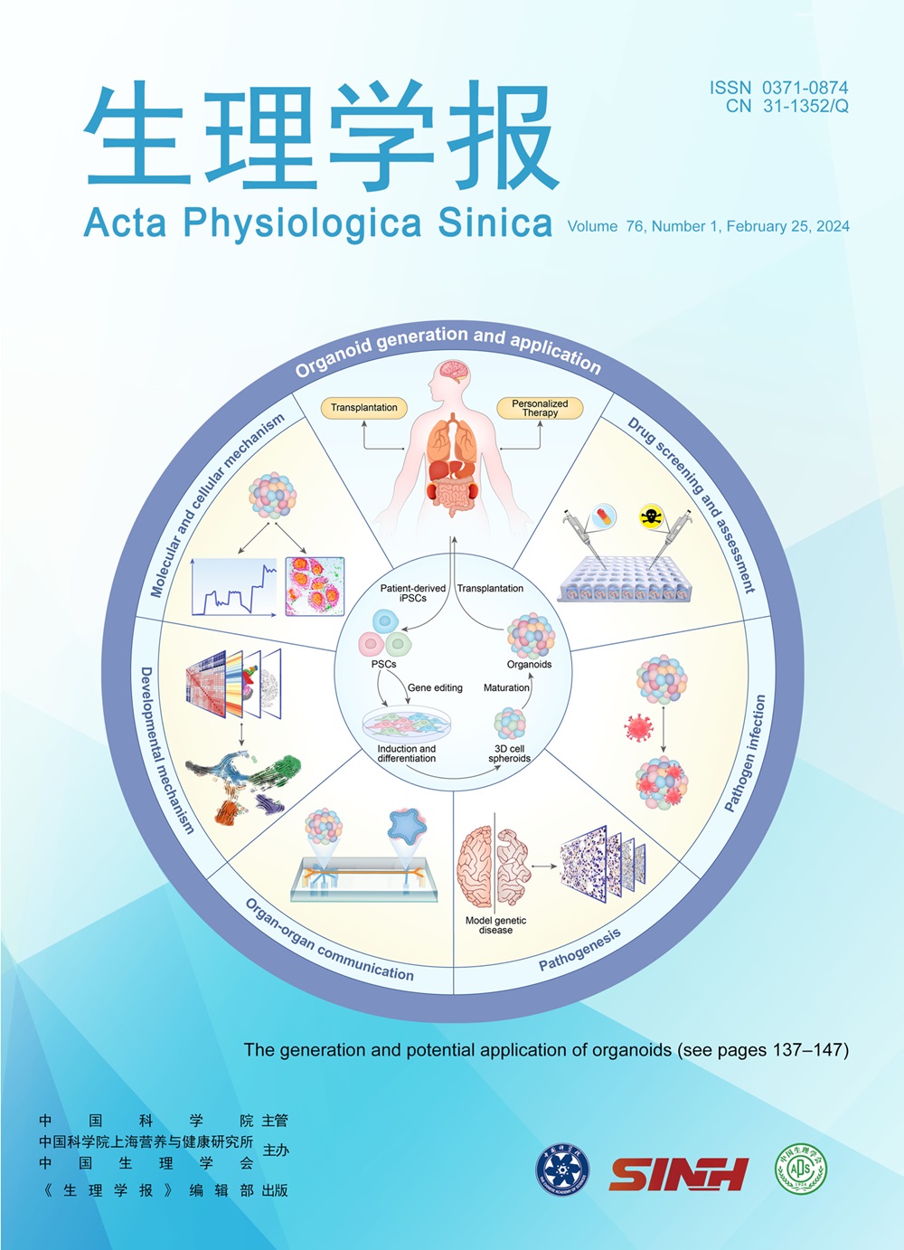
期刊介绍:
Acta Physiologica Sinica (APS) is sponsored by the Chinese Association for Physiological Sciences and Shanghai Institutes of Biological Sciences, Chinese Academy of Sciences (CAS), and is published bimonthly by the Science Press, China. APS publishes original research articles in the field of physiology as well as research contributions from other biomedical disciplines and proceedings of conferences and symposia of physiological sciences. Besides “Original Research Articles”, the journal also provides columns as “Brief Review”, “Rapid Communication”, “Experimental Technique”, and “Letter to the Editor”. Articles are published in either Chinese or English according to authors’ submission.