Kwang Ho Cho, Yuki Sugiyama, Genji Watanabe, Hidetomo Hirouchi, Gen Murakami, Jose Francisco Rodríguez-Vázquez, Shin-ichi Abe
{"title":"供应下唇的马氏神经分支再研究:对人类胎儿和捐赠的老年尸体的研究","authors":"Kwang Ho Cho, Yuki Sugiyama, Genji Watanabe, Hidetomo Hirouchi, Gen Murakami, Jose Francisco Rodríguez-Vázquez, Shin-ichi Abe","doi":"10.1007/s00276-024-03365-2","DOIUrl":null,"url":null,"abstract":"<h3 data-test=\"abstract-sub-heading\">Purpose</h3><p>Little information is known about the mentalis nerve course from the lower lip approximation margin (free margin) to the upper lip. Likewise, no difference in nerve distribution has been observed between the cutaneous and mucosal parts of the lip. Therefore, this study reexamined mentalis nerve morphology.</p><h3 data-test=\"abstract-sub-heading\">Methods</h3><p>For macroscopic observations, three fresh cadavers were dissected (one male and two females; aged 78–93). We also evaluated histological sections obtained from five donated elderly cadavers (two males and three females, aged 82–96 years) and 15 human fetuses (11–40 weeks or crown–rump length 80–372 mm). Immunohistochemical analysis for S100 protein and tyrosine hydroxylase was performed.</p><h3 data-test=\"abstract-sub-heading\">Results</h3><p>In both fetuses and adult cadavers, one to three nerve branches ran upward in the submucosal tissue from the mental foramen. Near the free margin of the lip, some branches passed through the orbicularis oris muscle layer toward the lip skin, whereas others followed a reversed J-shaped course along the free margin. Nerve twigs ran in parallel beneath the mucosa, whereas wavy nerve twigs attached to the basal lamina of the lip epidermis. The difference in nerve endings abruptly occurred at the skin–mucosal junction. Tyrosine hydroxylase-positive sympathetic nerve twigs surrounded arteries and formed a branch composed of S100-negative unmyelinated fibers.</p><h3 data-test=\"abstract-sub-heading\">Conclusion</h3><p>The lower lip skin was innervated by a perforating branch passing through the orbicularis oris muscle, that was different from the lip mucosa. A sudden change in the nerve ending configuration at the mucocutaneous junction seemed to develop postnatally.</p>","PeriodicalId":49296,"journal":{"name":"Surgical and Radiologic Anatomy","volume":"18 1","pages":""},"PeriodicalIF":1.2000,"publicationDate":"2024-04-29","publicationTypes":"Journal Article","fieldsOfStudy":null,"isOpenAccess":false,"openAccessPdf":"","citationCount":"0","resultStr":"{\"title\":\"Mentalis nerve branches supplying the lower lip revisited: a study of human fetuses and donated elderly cadavers\",\"authors\":\"Kwang Ho Cho, Yuki Sugiyama, Genji Watanabe, Hidetomo Hirouchi, Gen Murakami, Jose Francisco Rodríguez-Vázquez, Shin-ichi Abe\",\"doi\":\"10.1007/s00276-024-03365-2\",\"DOIUrl\":null,\"url\":null,\"abstract\":\"<h3 data-test=\\\"abstract-sub-heading\\\">Purpose</h3><p>Little information is known about the mentalis nerve course from the lower lip approximation margin (free margin) to the upper lip. Likewise, no difference in nerve distribution has been observed between the cutaneous and mucosal parts of the lip. Therefore, this study reexamined mentalis nerve morphology.</p><h3 data-test=\\\"abstract-sub-heading\\\">Methods</h3><p>For macroscopic observations, three fresh cadavers were dissected (one male and two females; aged 78–93). We also evaluated histological sections obtained from five donated elderly cadavers (two males and three females, aged 82–96 years) and 15 human fetuses (11–40 weeks or crown–rump length 80–372 mm). Immunohistochemical analysis for S100 protein and tyrosine hydroxylase was performed.</p><h3 data-test=\\\"abstract-sub-heading\\\">Results</h3><p>In both fetuses and adult cadavers, one to three nerve branches ran upward in the submucosal tissue from the mental foramen. Near the free margin of the lip, some branches passed through the orbicularis oris muscle layer toward the lip skin, whereas others followed a reversed J-shaped course along the free margin. Nerve twigs ran in parallel beneath the mucosa, whereas wavy nerve twigs attached to the basal lamina of the lip epidermis. The difference in nerve endings abruptly occurred at the skin–mucosal junction. Tyrosine hydroxylase-positive sympathetic nerve twigs surrounded arteries and formed a branch composed of S100-negative unmyelinated fibers.</p><h3 data-test=\\\"abstract-sub-heading\\\">Conclusion</h3><p>The lower lip skin was innervated by a perforating branch passing through the orbicularis oris muscle, that was different from the lip mucosa. A sudden change in the nerve ending configuration at the mucocutaneous junction seemed to develop postnatally.</p>\",\"PeriodicalId\":49296,\"journal\":{\"name\":\"Surgical and Radiologic Anatomy\",\"volume\":\"18 1\",\"pages\":\"\"},\"PeriodicalIF\":1.2000,\"publicationDate\":\"2024-04-29\",\"publicationTypes\":\"Journal Article\",\"fieldsOfStudy\":null,\"isOpenAccess\":false,\"openAccessPdf\":\"\",\"citationCount\":\"0\",\"resultStr\":null,\"platform\":\"Semanticscholar\",\"paperid\":null,\"PeriodicalName\":\"Surgical and Radiologic Anatomy\",\"FirstCategoryId\":\"3\",\"ListUrlMain\":\"https://doi.org/10.1007/s00276-024-03365-2\",\"RegionNum\":4,\"RegionCategory\":\"医学\",\"ArticlePicture\":[],\"TitleCN\":null,\"AbstractTextCN\":null,\"PMCID\":null,\"EPubDate\":\"\",\"PubModel\":\"\",\"JCR\":\"Q3\",\"JCRName\":\"ANATOMY & MORPHOLOGY\",\"Score\":null,\"Total\":0}","platform":"Semanticscholar","paperid":null,"PeriodicalName":"Surgical and Radiologic Anatomy","FirstCategoryId":"3","ListUrlMain":"https://doi.org/10.1007/s00276-024-03365-2","RegionNum":4,"RegionCategory":"医学","ArticlePicture":[],"TitleCN":null,"AbstractTextCN":null,"PMCID":null,"EPubDate":"","PubModel":"","JCR":"Q3","JCRName":"ANATOMY & MORPHOLOGY","Score":null,"Total":0}
Mentalis nerve branches supplying the lower lip revisited: a study of human fetuses and donated elderly cadavers
Purpose
Little information is known about the mentalis nerve course from the lower lip approximation margin (free margin) to the upper lip. Likewise, no difference in nerve distribution has been observed between the cutaneous and mucosal parts of the lip. Therefore, this study reexamined mentalis nerve morphology.
Methods
For macroscopic observations, three fresh cadavers were dissected (one male and two females; aged 78–93). We also evaluated histological sections obtained from five donated elderly cadavers (two males and three females, aged 82–96 years) and 15 human fetuses (11–40 weeks or crown–rump length 80–372 mm). Immunohistochemical analysis for S100 protein and tyrosine hydroxylase was performed.
Results
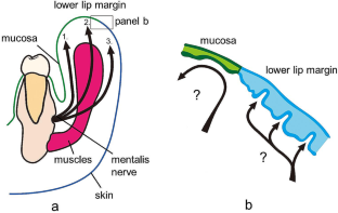
In both fetuses and adult cadavers, one to three nerve branches ran upward in the submucosal tissue from the mental foramen. Near the free margin of the lip, some branches passed through the orbicularis oris muscle layer toward the lip skin, whereas others followed a reversed J-shaped course along the free margin. Nerve twigs ran in parallel beneath the mucosa, whereas wavy nerve twigs attached to the basal lamina of the lip epidermis. The difference in nerve endings abruptly occurred at the skin–mucosal junction. Tyrosine hydroxylase-positive sympathetic nerve twigs surrounded arteries and formed a branch composed of S100-negative unmyelinated fibers.
Conclusion
The lower lip skin was innervated by a perforating branch passing through the orbicularis oris muscle, that was different from the lip mucosa. A sudden change in the nerve ending configuration at the mucocutaneous junction seemed to develop postnatally.
期刊介绍:
Anatomy is a morphological science which cannot fail to interest the clinician. The practical application of anatomical research to clinical problems necessitates special adaptation and selectivity in choosing from numerous international works. Although there is a tendency to believe that meaningful advances in anatomy are unlikely, constant revision is necessary. Surgical and Radiologic Anatomy, the first international journal of Clinical anatomy has been created in this spirit.
Its goal is to serve clinicians, regardless of speciality-physicians, surgeons, radiologists or other specialists-as an indispensable aid with which they can improve their knowledge of anatomy. Each issue includes: Original papers, review articles, articles on the anatomical bases of medical, surgical and radiological techniques, articles of normal radiologic anatomy, brief reviews of anatomical publications of clinical interest.
Particular attention is given to high quality illustrations, which are indispensable for a better understanding of anatomical problems.
Surgical and Radiologic Anatomy is a journal written by anatomists for clinicians with a special interest in anatomy.

 求助内容:
求助内容: 应助结果提醒方式:


