Jingjing Zhao, Zihui Li, Haixi Zhang, Tao Qin, Juan Zhao, Qiang Pei
{"title":"重组水蛭素通过调节 PAR-1-VEGF 抑制弥漫大 B 细胞淋巴瘤的血管生成","authors":"Jingjing Zhao, Zihui Li, Haixi Zhang, Tao Qin, Juan Zhao, Qiang Pei","doi":"10.1111/cbdd.14533","DOIUrl":null,"url":null,"abstract":"<p>Hirudin is one of the specific inhibitors of thrombin, which has been confirmed to have strong bioactivities, including inhibiting tumors. However, the function and mechanism of hirudin and protease-activated receptor 1 (PAR-1) in diffuse large B-cell lymphoma (DLBCL) have not been clear. Detecting the expression PAR-1 in DLBCL tissues and cells by RT-qPCR and IHC. Transfected sh-NC, sh-PAR-1, or pcDNA3.1-PAR-1 in DLBCL cells or processed DLBCL cells through added thrombin, Vorapaxar, Recombinant hirudin (RH), or Na<sub>2</sub>S<sub>2</sub>O<sub>4</sub> and co-culture with EA.hy926. And built DLBCL mice observed tumor growth. Detecting the expression of related genes by RT-qPCR, Western blot, IHC, and immunofluorescence, measured the cellular hypoxia with Hypoxyprobe-1 Kit, and estimated the cell inflammatory factors, proliferation, migration, invasion, and apoptosis by ELISA, CCK-8, flow cytometry, wound-healing and Transwell. Co-immunoprecipitation and pull-down measurement were used to verify the relationship. PAR-1 was highly expressed in DLBCL tissues and cells, especially in SUDHL2. Na<sub>2</sub>S<sub>2</sub>O<sub>4</sub> induced SUDHL2 hypoxia, and PAR-1 did not influence thrombin-activated hypoxia. PAR-1 could promote SUDHL2 proliferation, migration, and invasion, and it was unrelated to cellular hypoxia. PAR-1 promoted proliferation, migration, and angiogenesis of EA.hy926 or SUDHL2 through up-regulation vascular endothelial growth factor (VEGF). RH inhibited tumor growth, cell proliferation, and migration, promoted apoptosis of DLBCL, and inhibited angiogenesis by down-regulating PAR-1-VEGF. RH inhibits proliferation, migration, and angiogenesis of DLBCL cells by down-regulating PAR-1-VEGF.</p>","PeriodicalId":143,"journal":{"name":"Chemical Biology & Drug Design","volume":null,"pages":null},"PeriodicalIF":3.2000,"publicationDate":"2024-04-29","publicationTypes":"Journal Article","fieldsOfStudy":null,"isOpenAccess":false,"openAccessPdf":"","citationCount":"0","resultStr":"{\"title\":\"Recombinant hirudin suppresses angiogenesis of diffuse large B-cell lymphoma through regulation of the PAR-1-VEGF\",\"authors\":\"Jingjing Zhao, Zihui Li, Haixi Zhang, Tao Qin, Juan Zhao, Qiang Pei\",\"doi\":\"10.1111/cbdd.14533\",\"DOIUrl\":null,\"url\":null,\"abstract\":\"<p>Hirudin is one of the specific inhibitors of thrombin, which has been confirmed to have strong bioactivities, including inhibiting tumors. However, the function and mechanism of hirudin and protease-activated receptor 1 (PAR-1) in diffuse large B-cell lymphoma (DLBCL) have not been clear. Detecting the expression PAR-1 in DLBCL tissues and cells by RT-qPCR and IHC. Transfected sh-NC, sh-PAR-1, or pcDNA3.1-PAR-1 in DLBCL cells or processed DLBCL cells through added thrombin, Vorapaxar, Recombinant hirudin (RH), or Na<sub>2</sub>S<sub>2</sub>O<sub>4</sub> and co-culture with EA.hy926. And built DLBCL mice observed tumor growth. Detecting the expression of related genes by RT-qPCR, Western blot, IHC, and immunofluorescence, measured the cellular hypoxia with Hypoxyprobe-1 Kit, and estimated the cell inflammatory factors, proliferation, migration, invasion, and apoptosis by ELISA, CCK-8, flow cytometry, wound-healing and Transwell. Co-immunoprecipitation and pull-down measurement were used to verify the relationship. PAR-1 was highly expressed in DLBCL tissues and cells, especially in SUDHL2. Na<sub>2</sub>S<sub>2</sub>O<sub>4</sub> induced SUDHL2 hypoxia, and PAR-1 did not influence thrombin-activated hypoxia. PAR-1 could promote SUDHL2 proliferation, migration, and invasion, and it was unrelated to cellular hypoxia. PAR-1 promoted proliferation, migration, and angiogenesis of EA.hy926 or SUDHL2 through up-regulation vascular endothelial growth factor (VEGF). RH inhibited tumor growth, cell proliferation, and migration, promoted apoptosis of DLBCL, and inhibited angiogenesis by down-regulating PAR-1-VEGF. RH inhibits proliferation, migration, and angiogenesis of DLBCL cells by down-regulating PAR-1-VEGF.</p>\",\"PeriodicalId\":143,\"journal\":{\"name\":\"Chemical Biology & Drug Design\",\"volume\":null,\"pages\":null},\"PeriodicalIF\":3.2000,\"publicationDate\":\"2024-04-29\",\"publicationTypes\":\"Journal Article\",\"fieldsOfStudy\":null,\"isOpenAccess\":false,\"openAccessPdf\":\"\",\"citationCount\":\"0\",\"resultStr\":null,\"platform\":\"Semanticscholar\",\"paperid\":null,\"PeriodicalName\":\"Chemical Biology & Drug Design\",\"FirstCategoryId\":\"3\",\"ListUrlMain\":\"https://onlinelibrary.wiley.com/doi/10.1111/cbdd.14533\",\"RegionNum\":4,\"RegionCategory\":\"医学\",\"ArticlePicture\":[],\"TitleCN\":null,\"AbstractTextCN\":null,\"PMCID\":null,\"EPubDate\":\"\",\"PubModel\":\"\",\"JCR\":\"Q2\",\"JCRName\":\"BIOCHEMISTRY & MOLECULAR BIOLOGY\",\"Score\":null,\"Total\":0}","platform":"Semanticscholar","paperid":null,"PeriodicalName":"Chemical Biology & Drug Design","FirstCategoryId":"3","ListUrlMain":"https://onlinelibrary.wiley.com/doi/10.1111/cbdd.14533","RegionNum":4,"RegionCategory":"医学","ArticlePicture":[],"TitleCN":null,"AbstractTextCN":null,"PMCID":null,"EPubDate":"","PubModel":"","JCR":"Q2","JCRName":"BIOCHEMISTRY & MOLECULAR BIOLOGY","Score":null,"Total":0}
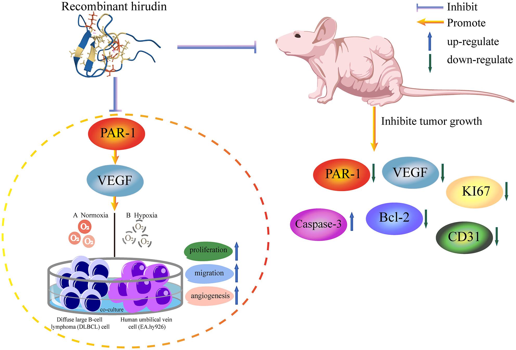
Recombinant hirudin suppresses angiogenesis of diffuse large B-cell lymphoma through regulation of the PAR-1-VEGF
Hirudin is one of the specific inhibitors of thrombin, which has been confirmed to have strong bioactivities, including inhibiting tumors. However, the function and mechanism of hirudin and protease-activated receptor 1 (PAR-1) in diffuse large B-cell lymphoma (DLBCL) have not been clear. Detecting the expression PAR-1 in DLBCL tissues and cells by RT-qPCR and IHC. Transfected sh-NC, sh-PAR-1, or pcDNA3.1-PAR-1 in DLBCL cells or processed DLBCL cells through added thrombin, Vorapaxar, Recombinant hirudin (RH), or Na2S2O4 and co-culture with EA.hy926. And built DLBCL mice observed tumor growth. Detecting the expression of related genes by RT-qPCR, Western blot, IHC, and immunofluorescence, measured the cellular hypoxia with Hypoxyprobe-1 Kit, and estimated the cell inflammatory factors, proliferation, migration, invasion, and apoptosis by ELISA, CCK-8, flow cytometry, wound-healing and Transwell. Co-immunoprecipitation and pull-down measurement were used to verify the relationship. PAR-1 was highly expressed in DLBCL tissues and cells, especially in SUDHL2. Na2S2O4 induced SUDHL2 hypoxia, and PAR-1 did not influence thrombin-activated hypoxia. PAR-1 could promote SUDHL2 proliferation, migration, and invasion, and it was unrelated to cellular hypoxia. PAR-1 promoted proliferation, migration, and angiogenesis of EA.hy926 or SUDHL2 through up-regulation vascular endothelial growth factor (VEGF). RH inhibited tumor growth, cell proliferation, and migration, promoted apoptosis of DLBCL, and inhibited angiogenesis by down-regulating PAR-1-VEGF. RH inhibits proliferation, migration, and angiogenesis of DLBCL cells by down-regulating PAR-1-VEGF.
期刊介绍:
Chemical Biology & Drug Design is a peer-reviewed scientific journal that is dedicated to the advancement of innovative science, technology and medicine with a focus on the multidisciplinary fields of chemical biology and drug design. It is the aim of Chemical Biology & Drug Design to capture significant research and drug discovery that highlights new concepts, insight and new findings within the scope of chemical biology and drug design.

 求助内容:
求助内容: 应助结果提醒方式:


