{"title":"微生物红蛋白中视网膜质子化希夫碱的固态核磁共振成像","authors":"","doi":"10.1016/j.mrl.2024.200132","DOIUrl":null,"url":null,"abstract":"<div><p>Rhodopsin is a seven-helical transmembrane protein with a retinal chromophore covalently bound to a conserved lysine in helix G via a retinal protonated Schiff base (RPSB). Microbial rhodopsins absorb light through chromophore and play a fundamental role in optogenetics. Numerous microbial rhodopsins have been discovered, contributing to diverse functions and colors. Solid-state NMR spectroscopy has been instrumental in elucidating the conformation of chromophores and the three-dimensional structure of microbial rhodopsins. This review focuses on the <sup>15</sup>N chemical shift values of RPSB and summarizes recent progress in the field. We displayed the correlation between the <sup>15</sup>N isotropic chemical shift values of RPSB and the maximum absorption wavelength of rhodopsin using solid-state NMR spectroscopy.</p></div>","PeriodicalId":93594,"journal":{"name":"Magnetic Resonance Letters","volume":"4 3","pages":"Article 200132"},"PeriodicalIF":0.0000,"publicationDate":"2024-08-01","publicationTypes":"Journal Article","fieldsOfStudy":null,"isOpenAccess":false,"openAccessPdf":"https://www.sciencedirect.com/science/article/pii/S2772516224000391/pdfft?md5=498b14a4e213885e68caec1c0a74709f&pid=1-s2.0-S2772516224000391-main.pdf","citationCount":"0","resultStr":"{\"title\":\"Solid-state NMR of the retinal protonated Schiff base in microbial rhodopsins\",\"authors\":\"\",\"doi\":\"10.1016/j.mrl.2024.200132\",\"DOIUrl\":null,\"url\":null,\"abstract\":\"<div><p>Rhodopsin is a seven-helical transmembrane protein with a retinal chromophore covalently bound to a conserved lysine in helix G via a retinal protonated Schiff base (RPSB). Microbial rhodopsins absorb light through chromophore and play a fundamental role in optogenetics. Numerous microbial rhodopsins have been discovered, contributing to diverse functions and colors. Solid-state NMR spectroscopy has been instrumental in elucidating the conformation of chromophores and the three-dimensional structure of microbial rhodopsins. This review focuses on the <sup>15</sup>N chemical shift values of RPSB and summarizes recent progress in the field. We displayed the correlation between the <sup>15</sup>N isotropic chemical shift values of RPSB and the maximum absorption wavelength of rhodopsin using solid-state NMR spectroscopy.</p></div>\",\"PeriodicalId\":93594,\"journal\":{\"name\":\"Magnetic Resonance Letters\",\"volume\":\"4 3\",\"pages\":\"Article 200132\"},\"PeriodicalIF\":0.0000,\"publicationDate\":\"2024-08-01\",\"publicationTypes\":\"Journal Article\",\"fieldsOfStudy\":null,\"isOpenAccess\":false,\"openAccessPdf\":\"https://www.sciencedirect.com/science/article/pii/S2772516224000391/pdfft?md5=498b14a4e213885e68caec1c0a74709f&pid=1-s2.0-S2772516224000391-main.pdf\",\"citationCount\":\"0\",\"resultStr\":null,\"platform\":\"Semanticscholar\",\"paperid\":null,\"PeriodicalName\":\"Magnetic Resonance Letters\",\"FirstCategoryId\":\"1085\",\"ListUrlMain\":\"https://www.sciencedirect.com/science/article/pii/S2772516224000391\",\"RegionNum\":0,\"RegionCategory\":null,\"ArticlePicture\":[],\"TitleCN\":null,\"AbstractTextCN\":null,\"PMCID\":null,\"EPubDate\":\"\",\"PubModel\":\"\",\"JCR\":\"\",\"JCRName\":\"\",\"Score\":null,\"Total\":0}","platform":"Semanticscholar","paperid":null,"PeriodicalName":"Magnetic Resonance Letters","FirstCategoryId":"1085","ListUrlMain":"https://www.sciencedirect.com/science/article/pii/S2772516224000391","RegionNum":0,"RegionCategory":null,"ArticlePicture":[],"TitleCN":null,"AbstractTextCN":null,"PMCID":null,"EPubDate":"","PubModel":"","JCR":"","JCRName":"","Score":null,"Total":0}
Solid-state NMR of the retinal protonated Schiff base in microbial rhodopsins
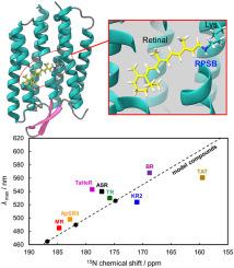
Rhodopsin is a seven-helical transmembrane protein with a retinal chromophore covalently bound to a conserved lysine in helix G via a retinal protonated Schiff base (RPSB). Microbial rhodopsins absorb light through chromophore and play a fundamental role in optogenetics. Numerous microbial rhodopsins have been discovered, contributing to diverse functions and colors. Solid-state NMR spectroscopy has been instrumental in elucidating the conformation of chromophores and the three-dimensional structure of microbial rhodopsins. This review focuses on the 15N chemical shift values of RPSB and summarizes recent progress in the field. We displayed the correlation between the 15N isotropic chemical shift values of RPSB and the maximum absorption wavelength of rhodopsin using solid-state NMR spectroscopy.

 求助内容:
求助内容: 应助结果提醒方式:


