Pierleone Lucatelli, Simone Ciaglia, Bianca Rocco, Gianluca De Rubeis, Guido Bolognesi, Elio Damato, Mario Corona, Pier Giorgio Nardis, Alessandro Cannavale, Paolo Ricci, Carlo Catalano
{"title":"二维灌注血管造影可直接观察 b-TACE 期间肝细胞癌的血流再分布情况","authors":"Pierleone Lucatelli, Simone Ciaglia, Bianca Rocco, Gianluca De Rubeis, Guido Bolognesi, Elio Damato, Mario Corona, Pier Giorgio Nardis, Alessandro Cannavale, Paolo Ricci, Carlo Catalano","doi":"10.1007/s11547-024-01816-9","DOIUrl":null,"url":null,"abstract":"<h3 data-test=\"abstract-sub-heading\">Objectives</h3><p>To demonstrate in vivo redistribution of the blood flow towards HCC’s lesions by utilizing two-dimensional perfusion angiography in b-TACE procedures.</p><h3 data-test=\"abstract-sub-heading\">Material and methods</h3><p>In total, 30 patients with 35 HCC nodules treated in the period between January 2019 and November 2021. For each patient, a post-processing software leading to a two-dimensional perfusion angiography was applied on each angiography performed via balloon microcatheter, before and after inflation. On the colour map obtained, reflecting the evolution of contrast intensity change over time, five regions of interests (ROIs) were assessed: one on the tumour (ROI-t), two in the immediate peritumoural healthy liver parenchyma (ROI-ihl) and two in the peripheral healthy liver parenchyma (ROI-phl). The results have been interpreted with a novel in silico model that simulates the hemodynamics of the hepatic arterial system.</p><h3 data-test=\"abstract-sub-heading\">Results</h3><p>Among the ROIs drawn inside the same segment of target lesion, the time-to-peak of the ROI-t and of the ROI-ihl have a significantly higher mean value when the balloon was inflated compared with the ROIs obtained with deflated balloon (10.33 ± 3.66 s vs 8.87 ± 2.60 s (<i>p</i> = 0.015) for ROI-t; 10.50 ± 3.65 s vs 9.23 ± 2.70 s (<i>p</i> = 0.047) for ROI-ihl). The in silico model prediction time-to-peak delays when balloon was inflated, match with those observed in vivo. The numerical flow analysis shows how time-to-peak delays are caused by the obstruction of the balloon-occluded artery and the opening of intra-hepatic collateral.</p><h3 data-test=\"abstract-sub-heading\">Conclusion</h3><p>The measurements identify predictively the flow redistribution in the hepatic arteries during b-TACE, supporting a proper positioning of the balloon microcatheter.</p>","PeriodicalId":501689,"journal":{"name":"La radiologia medica","volume":"50 1","pages":""},"PeriodicalIF":0.0000,"publicationDate":"2024-04-18","publicationTypes":"Journal Article","fieldsOfStudy":null,"isOpenAccess":false,"openAccessPdf":"","citationCount":"0","resultStr":"{\"title\":\"Two-dimensional perfusion angiography permits direct visualization of redistribution of flow in hepatocellular carcinoma during b-TACE\",\"authors\":\"Pierleone Lucatelli, Simone Ciaglia, Bianca Rocco, Gianluca De Rubeis, Guido Bolognesi, Elio Damato, Mario Corona, Pier Giorgio Nardis, Alessandro Cannavale, Paolo Ricci, Carlo Catalano\",\"doi\":\"10.1007/s11547-024-01816-9\",\"DOIUrl\":null,\"url\":null,\"abstract\":\"<h3 data-test=\\\"abstract-sub-heading\\\">Objectives</h3><p>To demonstrate in vivo redistribution of the blood flow towards HCC’s lesions by utilizing two-dimensional perfusion angiography in b-TACE procedures.</p><h3 data-test=\\\"abstract-sub-heading\\\">Material and methods</h3><p>In total, 30 patients with 35 HCC nodules treated in the period between January 2019 and November 2021. For each patient, a post-processing software leading to a two-dimensional perfusion angiography was applied on each angiography performed via balloon microcatheter, before and after inflation. On the colour map obtained, reflecting the evolution of contrast intensity change over time, five regions of interests (ROIs) were assessed: one on the tumour (ROI-t), two in the immediate peritumoural healthy liver parenchyma (ROI-ihl) and two in the peripheral healthy liver parenchyma (ROI-phl). The results have been interpreted with a novel in silico model that simulates the hemodynamics of the hepatic arterial system.</p><h3 data-test=\\\"abstract-sub-heading\\\">Results</h3><p>Among the ROIs drawn inside the same segment of target lesion, the time-to-peak of the ROI-t and of the ROI-ihl have a significantly higher mean value when the balloon was inflated compared with the ROIs obtained with deflated balloon (10.33 ± 3.66 s vs 8.87 ± 2.60 s (<i>p</i> = 0.015) for ROI-t; 10.50 ± 3.65 s vs 9.23 ± 2.70 s (<i>p</i> = 0.047) for ROI-ihl). The in silico model prediction time-to-peak delays when balloon was inflated, match with those observed in vivo. The numerical flow analysis shows how time-to-peak delays are caused by the obstruction of the balloon-occluded artery and the opening of intra-hepatic collateral.</p><h3 data-test=\\\"abstract-sub-heading\\\">Conclusion</h3><p>The measurements identify predictively the flow redistribution in the hepatic arteries during b-TACE, supporting a proper positioning of the balloon microcatheter.</p>\",\"PeriodicalId\":501689,\"journal\":{\"name\":\"La radiologia medica\",\"volume\":\"50 1\",\"pages\":\"\"},\"PeriodicalIF\":0.0000,\"publicationDate\":\"2024-04-18\",\"publicationTypes\":\"Journal Article\",\"fieldsOfStudy\":null,\"isOpenAccess\":false,\"openAccessPdf\":\"\",\"citationCount\":\"0\",\"resultStr\":null,\"platform\":\"Semanticscholar\",\"paperid\":null,\"PeriodicalName\":\"La radiologia medica\",\"FirstCategoryId\":\"1085\",\"ListUrlMain\":\"https://doi.org/10.1007/s11547-024-01816-9\",\"RegionNum\":0,\"RegionCategory\":null,\"ArticlePicture\":[],\"TitleCN\":null,\"AbstractTextCN\":null,\"PMCID\":null,\"EPubDate\":\"\",\"PubModel\":\"\",\"JCR\":\"\",\"JCRName\":\"\",\"Score\":null,\"Total\":0}","platform":"Semanticscholar","paperid":null,"PeriodicalName":"La radiologia medica","FirstCategoryId":"1085","ListUrlMain":"https://doi.org/10.1007/s11547-024-01816-9","RegionNum":0,"RegionCategory":null,"ArticlePicture":[],"TitleCN":null,"AbstractTextCN":null,"PMCID":null,"EPubDate":"","PubModel":"","JCR":"","JCRName":"","Score":null,"Total":0}
Two-dimensional perfusion angiography permits direct visualization of redistribution of flow in hepatocellular carcinoma during b-TACE
Objectives
To demonstrate in vivo redistribution of the blood flow towards HCC’s lesions by utilizing two-dimensional perfusion angiography in b-TACE procedures.
Material and methods
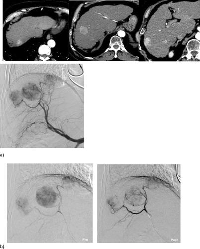
In total, 30 patients with 35 HCC nodules treated in the period between January 2019 and November 2021. For each patient, a post-processing software leading to a two-dimensional perfusion angiography was applied on each angiography performed via balloon microcatheter, before and after inflation. On the colour map obtained, reflecting the evolution of contrast intensity change over time, five regions of interests (ROIs) were assessed: one on the tumour (ROI-t), two in the immediate peritumoural healthy liver parenchyma (ROI-ihl) and two in the peripheral healthy liver parenchyma (ROI-phl). The results have been interpreted with a novel in silico model that simulates the hemodynamics of the hepatic arterial system.
Results
Among the ROIs drawn inside the same segment of target lesion, the time-to-peak of the ROI-t and of the ROI-ihl have a significantly higher mean value when the balloon was inflated compared with the ROIs obtained with deflated balloon (10.33 ± 3.66 s vs 8.87 ± 2.60 s (p = 0.015) for ROI-t; 10.50 ± 3.65 s vs 9.23 ± 2.70 s (p = 0.047) for ROI-ihl). The in silico model prediction time-to-peak delays when balloon was inflated, match with those observed in vivo. The numerical flow analysis shows how time-to-peak delays are caused by the obstruction of the balloon-occluded artery and the opening of intra-hepatic collateral.
Conclusion
The measurements identify predictively the flow redistribution in the hepatic arteries during b-TACE, supporting a proper positioning of the balloon microcatheter.

 求助内容:
求助内容: 应助结果提醒方式:


