Nicholas A. Waring, Alexander Chern, Brandon J. Vilarello, Yew Song Cheng, Chaoqun Zhou, Jeffrey H. Lang, Elizabeth S. Olson, Hideko Heidi Nakajima
{"title":"将汉普郡绵羊作为人工耳蜗植入的大型动物模型","authors":"Nicholas A. Waring, Alexander Chern, Brandon J. Vilarello, Yew Song Cheng, Chaoqun Zhou, Jeffrey H. Lang, Elizabeth S. Olson, Hideko Heidi Nakajima","doi":"10.1007/s10162-024-00946-1","DOIUrl":null,"url":null,"abstract":"<h3 data-test=\"abstract-sub-heading\">Background</h3><p>Sheep have been proposed as a large-animal model for studying cochlear implantation. However, prior sheep studies report that the facial nerve (FN) obscures the round window membrane (RWM), requiring FN sacrifice or a retrofacial opening to access the middle-ear cavity posterior to the FN for cochlear implantation. We investigated surgical access to the RWM in Hampshire sheep compared to Suffolk-Dorset sheep and the feasibility of Hampshire sheep for cochlear implantation via a facial recess approach.</p><h3 data-test=\"abstract-sub-heading\">Methods</h3><p>Sixteen temporal bones from cadaveric sheep heads (ten Hampshire and six Suffolk-Dorset) were dissected to gain surgical access to the RWM via an extended facial recess approach. RWM visibility was graded using St. Thomas’ Hospital (STH) classification. Cochlear implant (CI) electrode array insertion was performed in two Hampshire specimens. Micro-CT scans were obtained for each temporal bone, with confirmation of appropriate electrode array placement and segmentation of the inner ear structures.</p><h3 data-test=\"abstract-sub-heading\">Results</h3><p>Visibility of the RWM on average was 83% in Hampshire specimens and 59% in Suffolk-Dorset specimens (<i>p</i> = 0.0262). Hampshire RWM visibility was Type I (100% visibility) for three specimens and Type IIa (> 50% visibility) for seven specimens. Suffolk-Dorset RWM visibility was Type IIa for four specimens and Type IIb (< 50% visibility) for two specimens. FN appeared to course more anterolaterally in Suffolk-Dorset specimens. Micro-CT confirmed appropriate CI electrode array placement in the scala tympani without apparent basilar membrane rupture.</p><h3 data-test=\"abstract-sub-heading\">Conclusions</h3><p>Hampshire sheep appear to be a suitable large-animal model for CI electrode insertion via an extended facial recess approach without sacrificing the FN. In this small sample, Hampshire specimens had improved RWM visibility compared to Suffolk-Dorset. Thus, Hampshire sheep may be superior to other breeds for ease of cochlear implantation, with FN and facial recess anatomy more similar to humans.</p>","PeriodicalId":17236,"journal":{"name":"Journal of the Association for Research in Otolaryngology","volume":"5 1","pages":""},"PeriodicalIF":0.0000,"publicationDate":"2024-04-15","publicationTypes":"Journal Article","fieldsOfStudy":null,"isOpenAccess":false,"openAccessPdf":"","citationCount":"0","resultStr":"{\"title\":\"Hampshire Sheep as a Large-Animal Model for Cochlear Implantation\",\"authors\":\"Nicholas A. Waring, Alexander Chern, Brandon J. Vilarello, Yew Song Cheng, Chaoqun Zhou, Jeffrey H. Lang, Elizabeth S. Olson, Hideko Heidi Nakajima\",\"doi\":\"10.1007/s10162-024-00946-1\",\"DOIUrl\":null,\"url\":null,\"abstract\":\"<h3 data-test=\\\"abstract-sub-heading\\\">Background</h3><p>Sheep have been proposed as a large-animal model for studying cochlear implantation. However, prior sheep studies report that the facial nerve (FN) obscures the round window membrane (RWM), requiring FN sacrifice or a retrofacial opening to access the middle-ear cavity posterior to the FN for cochlear implantation. We investigated surgical access to the RWM in Hampshire sheep compared to Suffolk-Dorset sheep and the feasibility of Hampshire sheep for cochlear implantation via a facial recess approach.</p><h3 data-test=\\\"abstract-sub-heading\\\">Methods</h3><p>Sixteen temporal bones from cadaveric sheep heads (ten Hampshire and six Suffolk-Dorset) were dissected to gain surgical access to the RWM via an extended facial recess approach. RWM visibility was graded using St. Thomas’ Hospital (STH) classification. Cochlear implant (CI) electrode array insertion was performed in two Hampshire specimens. Micro-CT scans were obtained for each temporal bone, with confirmation of appropriate electrode array placement and segmentation of the inner ear structures.</p><h3 data-test=\\\"abstract-sub-heading\\\">Results</h3><p>Visibility of the RWM on average was 83% in Hampshire specimens and 59% in Suffolk-Dorset specimens (<i>p</i> = 0.0262). Hampshire RWM visibility was Type I (100% visibility) for three specimens and Type IIa (> 50% visibility) for seven specimens. Suffolk-Dorset RWM visibility was Type IIa for four specimens and Type IIb (< 50% visibility) for two specimens. FN appeared to course more anterolaterally in Suffolk-Dorset specimens. Micro-CT confirmed appropriate CI electrode array placement in the scala tympani without apparent basilar membrane rupture.</p><h3 data-test=\\\"abstract-sub-heading\\\">Conclusions</h3><p>Hampshire sheep appear to be a suitable large-animal model for CI electrode insertion via an extended facial recess approach without sacrificing the FN. In this small sample, Hampshire specimens had improved RWM visibility compared to Suffolk-Dorset. Thus, Hampshire sheep may be superior to other breeds for ease of cochlear implantation, with FN and facial recess anatomy more similar to humans.</p>\",\"PeriodicalId\":17236,\"journal\":{\"name\":\"Journal of the Association for Research in Otolaryngology\",\"volume\":\"5 1\",\"pages\":\"\"},\"PeriodicalIF\":0.0000,\"publicationDate\":\"2024-04-15\",\"publicationTypes\":\"Journal Article\",\"fieldsOfStudy\":null,\"isOpenAccess\":false,\"openAccessPdf\":\"\",\"citationCount\":\"0\",\"resultStr\":null,\"platform\":\"Semanticscholar\",\"paperid\":null,\"PeriodicalName\":\"Journal of the Association for Research in Otolaryngology\",\"FirstCategoryId\":\"1085\",\"ListUrlMain\":\"https://doi.org/10.1007/s10162-024-00946-1\",\"RegionNum\":0,\"RegionCategory\":null,\"ArticlePicture\":[],\"TitleCN\":null,\"AbstractTextCN\":null,\"PMCID\":null,\"EPubDate\":\"\",\"PubModel\":\"\",\"JCR\":\"\",\"JCRName\":\"\",\"Score\":null,\"Total\":0}","platform":"Semanticscholar","paperid":null,"PeriodicalName":"Journal of the Association for Research in Otolaryngology","FirstCategoryId":"1085","ListUrlMain":"https://doi.org/10.1007/s10162-024-00946-1","RegionNum":0,"RegionCategory":null,"ArticlePicture":[],"TitleCN":null,"AbstractTextCN":null,"PMCID":null,"EPubDate":"","PubModel":"","JCR":"","JCRName":"","Score":null,"Total":0}
Hampshire Sheep as a Large-Animal Model for Cochlear Implantation
Background
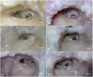
Sheep have been proposed as a large-animal model for studying cochlear implantation. However, prior sheep studies report that the facial nerve (FN) obscures the round window membrane (RWM), requiring FN sacrifice or a retrofacial opening to access the middle-ear cavity posterior to the FN for cochlear implantation. We investigated surgical access to the RWM in Hampshire sheep compared to Suffolk-Dorset sheep and the feasibility of Hampshire sheep for cochlear implantation via a facial recess approach.
Methods
Sixteen temporal bones from cadaveric sheep heads (ten Hampshire and six Suffolk-Dorset) were dissected to gain surgical access to the RWM via an extended facial recess approach. RWM visibility was graded using St. Thomas’ Hospital (STH) classification. Cochlear implant (CI) electrode array insertion was performed in two Hampshire specimens. Micro-CT scans were obtained for each temporal bone, with confirmation of appropriate electrode array placement and segmentation of the inner ear structures.
Results
Visibility of the RWM on average was 83% in Hampshire specimens and 59% in Suffolk-Dorset specimens (p = 0.0262). Hampshire RWM visibility was Type I (100% visibility) for three specimens and Type IIa (> 50% visibility) for seven specimens. Suffolk-Dorset RWM visibility was Type IIa for four specimens and Type IIb (< 50% visibility) for two specimens. FN appeared to course more anterolaterally in Suffolk-Dorset specimens. Micro-CT confirmed appropriate CI electrode array placement in the scala tympani without apparent basilar membrane rupture.
Conclusions
Hampshire sheep appear to be a suitable large-animal model for CI electrode insertion via an extended facial recess approach without sacrificing the FN. In this small sample, Hampshire specimens had improved RWM visibility compared to Suffolk-Dorset. Thus, Hampshire sheep may be superior to other breeds for ease of cochlear implantation, with FN and facial recess anatomy more similar to humans.

 求助内容:
求助内容: 应助结果提醒方式:


