通过高角度分辨率弥散成像对肾脏进行弥散牵引成像

Surendra Maharjan , Jie Chen , Adrienne Gaughan , Neal X. Chen , Nian Wang
{"title":"通过高角度分辨率弥散成像对肾脏进行弥散牵引成像","authors":"Surendra Maharjan ,&nbsp;Jie Chen ,&nbsp;Adrienne Gaughan ,&nbsp;Neal X. Chen ,&nbsp;Nian Wang","doi":"10.1016/j.mrl.2024.200117","DOIUrl":null,"url":null,"abstract":"<div><div>Diffusion magnetic resonance imaging (MRI) has been utilized to probe the renal microstructures but investigating the three-dimensional (3D) tubular network still presents significant challenges due to the complicated architecture of kidney. This study aims to assess whether high angular resolution diffusion imaging (HARDI) could improve the reconstruction of 3D tubular architectures. Kidneys from both mice and rats were imaged using 3D diffusion-weighted pulse sequences at 9.4 T. Five healthy mouse kidneys were scanned at an isotropic spatial resolution of 40 μm, with a <em>b</em> value of 1500 s/mm<sup>2</sup> across 46 diffusion encoding directions. The study employed diffusion tensor imaging (DTI) and generalized Q-sampling imaging (GQI) to examine the tubular orientation distributions and tractography, validated by conventional histology. Fractional anisotropy (FA) and mean diffusivity (MD) were quantified and compared among the inner medullar (IM), outer medullar (OM), and cortex (CO) at different angular resolutions. FA values, estimated with 6 diffusion-weighted images (DWIs), were significantly overestimated by 49.9% (<em>p</em> &lt; 0.001) in IM, 179.4% (<em>p</em> &lt; 0.001) in OM, and 225.5% (<em>p</em> &lt; 0.001) in CO, compared to using 46 DWIs. In contrast, MD exhibited less variations to angular resolution variations (3.4% in IM, 4.2% in OM, and 4.6% in CO). Both DTI and GQI at high angular resolution successfully traced renal tubular structures throughout the kidney, with GQI demonstrating superior performance in generating more continuous tracts. Furthermore, disrupted renal tubule structures were observed in a chronic kidney disease (CKD) rat model. HARDI, especially when combined with the GQI approach, holds promise in tracking complicated 3D tubule architectures and may serve as a potent tool for kidney disease research.</div></div>","PeriodicalId":93594,"journal":{"name":"Magnetic Resonance Letters","volume":"4 4","pages":"Article 200117"},"PeriodicalIF":0.0000,"publicationDate":"2024-11-01","publicationTypes":"Journal Article","fieldsOfStudy":null,"isOpenAccess":false,"openAccessPdf":"","citationCount":"0","resultStr":"{\"title\":\"Diffusion tractography of kidney by high angular resolution diffusion imaging\",\"authors\":\"Surendra Maharjan ,&nbsp;Jie Chen ,&nbsp;Adrienne Gaughan ,&nbsp;Neal X. Chen ,&nbsp;Nian Wang\",\"doi\":\"10.1016/j.mrl.2024.200117\",\"DOIUrl\":null,\"url\":null,\"abstract\":\"<div><div>Diffusion magnetic resonance imaging (MRI) has been utilized to probe the renal microstructures but investigating the three-dimensional (3D) tubular network still presents significant challenges due to the complicated architecture of kidney. This study aims to assess whether high angular resolution diffusion imaging (HARDI) could improve the reconstruction of 3D tubular architectures. Kidneys from both mice and rats were imaged using 3D diffusion-weighted pulse sequences at 9.4 T. Five healthy mouse kidneys were scanned at an isotropic spatial resolution of 40 μm, with a <em>b</em> value of 1500 s/mm<sup>2</sup> across 46 diffusion encoding directions. The study employed diffusion tensor imaging (DTI) and generalized Q-sampling imaging (GQI) to examine the tubular orientation distributions and tractography, validated by conventional histology. Fractional anisotropy (FA) and mean diffusivity (MD) were quantified and compared among the inner medullar (IM), outer medullar (OM), and cortex (CO) at different angular resolutions. FA values, estimated with 6 diffusion-weighted images (DWIs), were significantly overestimated by 49.9% (<em>p</em> &lt; 0.001) in IM, 179.4% (<em>p</em> &lt; 0.001) in OM, and 225.5% (<em>p</em> &lt; 0.001) in CO, compared to using 46 DWIs. In contrast, MD exhibited less variations to angular resolution variations (3.4% in IM, 4.2% in OM, and 4.6% in CO). Both DTI and GQI at high angular resolution successfully traced renal tubular structures throughout the kidney, with GQI demonstrating superior performance in generating more continuous tracts. Furthermore, disrupted renal tubule structures were observed in a chronic kidney disease (CKD) rat model. HARDI, especially when combined with the GQI approach, holds promise in tracking complicated 3D tubule architectures and may serve as a potent tool for kidney disease research.</div></div>\",\"PeriodicalId\":93594,\"journal\":{\"name\":\"Magnetic Resonance Letters\",\"volume\":\"4 4\",\"pages\":\"Article 200117\"},\"PeriodicalIF\":0.0000,\"publicationDate\":\"2024-11-01\",\"publicationTypes\":\"Journal Article\",\"fieldsOfStudy\":null,\"isOpenAccess\":false,\"openAccessPdf\":\"\",\"citationCount\":\"0\",\"resultStr\":null,\"platform\":\"Semanticscholar\",\"paperid\":null,\"PeriodicalName\":\"Magnetic Resonance Letters\",\"FirstCategoryId\":\"1085\",\"ListUrlMain\":\"https://www.sciencedirect.com/science/article/pii/S277251622400024X\",\"RegionNum\":0,\"RegionCategory\":null,\"ArticlePicture\":[],\"TitleCN\":null,\"AbstractTextCN\":null,\"PMCID\":null,\"EPubDate\":\"\",\"PubModel\":\"\",\"JCR\":\"\",\"JCRName\":\"\",\"Score\":null,\"Total\":0}","platform":"Semanticscholar","paperid":null,"PeriodicalName":"Magnetic Resonance Letters","FirstCategoryId":"1085","ListUrlMain":"https://www.sciencedirect.com/science/article/pii/S277251622400024X","RegionNum":0,"RegionCategory":null,"ArticlePicture":[],"TitleCN":null,"AbstractTextCN":null,"PMCID":null,"EPubDate":"","PubModel":"","JCR":"","JCRName":"","Score":null,"Total":0}
引用次数: 0

摘要

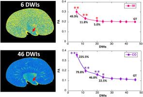
扩散磁共振成像(MRI)已被用于探测肾脏的微观结构,但由于肾脏结构复杂,研究三维(3D)肾小管网络仍面临巨大挑战。本研究旨在评估高角度分辨率弥散成像(HARDI)能否改善三维肾小管结构的重建。五只健康小鼠的肾脏在各向同性空间分辨率为 40 μm、b 值为 1500 s/mm2 的 46 个扩散编码方向上进行了扫描。研究采用了弥散张量成像(DTI)和广义 Q 采样成像(GQI)来检测肾小管的方向分布和束学,并通过传统组织学进行验证。在不同角度分辨率下,对内髓质(IM)、外髓质(OM)和皮质(CO)的分数各向异性(FA)和平均扩散率(MD)进行了量化和比较。与使用 46 张扩散加权图像(DWIs)相比,使用 6 张扩散加权图像(DWIs)估算的 FA 值在内髓被明显高估了 49.9% (p < 0.001),在外髓被高估了 179.4% (p < 0.001),在 CO 被高估了 225.5% (p < 0.001)。相比之下,MD 对角度分辨率变化的影响较小(IM 为 3.4%,OM 为 4.2%,CO 为 4.6%)。高角度分辨率下的 DTI 和 GQI 都能成功追踪整个肾脏的肾小管结构,而 GQI 在生成更连续的肾小管方面表现更优。此外,在慢性肾病(CKD)大鼠模型中也观察到了肾小管结构的破坏。HARDI,尤其是与 GQI 方法相结合时,有望追踪复杂的三维肾小管结构,并可作为肾脏疾病研究的有力工具。
本文章由计算机程序翻译,如有差异,请以英文原文为准。

Diffusion tractography of kidney by high angular resolution diffusion imaging

Diffusion tractography of kidney by high angular resolution diffusion imaging
Diffusion magnetic resonance imaging (MRI) has been utilized to probe the renal microstructures but investigating the three-dimensional (3D) tubular network still presents significant challenges due to the complicated architecture of kidney. This study aims to assess whether high angular resolution diffusion imaging (HARDI) could improve the reconstruction of 3D tubular architectures. Kidneys from both mice and rats were imaged using 3D diffusion-weighted pulse sequences at 9.4 T. Five healthy mouse kidneys were scanned at an isotropic spatial resolution of 40 μm, with a b value of 1500 s/mm2 across 46 diffusion encoding directions. The study employed diffusion tensor imaging (DTI) and generalized Q-sampling imaging (GQI) to examine the tubular orientation distributions and tractography, validated by conventional histology. Fractional anisotropy (FA) and mean diffusivity (MD) were quantified and compared among the inner medullar (IM), outer medullar (OM), and cortex (CO) at different angular resolutions. FA values, estimated with 6 diffusion-weighted images (DWIs), were significantly overestimated by 49.9% (p < 0.001) in IM, 179.4% (p < 0.001) in OM, and 225.5% (p < 0.001) in CO, compared to using 46 DWIs. In contrast, MD exhibited less variations to angular resolution variations (3.4% in IM, 4.2% in OM, and 4.6% in CO). Both DTI and GQI at high angular resolution successfully traced renal tubular structures throughout the kidney, with GQI demonstrating superior performance in generating more continuous tracts. Furthermore, disrupted renal tubule structures were observed in a chronic kidney disease (CKD) rat model. HARDI, especially when combined with the GQI approach, holds promise in tracking complicated 3D tubule architectures and may serve as a potent tool for kidney disease research.
求助全文
通过发布文献求助,成功后即可免费获取论文全文。 去求助
来源期刊
Magnetic Resonance Letters
Magnetic Resonance Letters Analytical Chemistry, Spectroscopy, Radiology and Imaging, Biochemistry, Genetics and Molecular Biology (General)
自引率
0.00%
发文量
0
×
引用
GB/T 7714-2015
复制
MLA
复制
APA
复制
导出至
BibTeX EndNote RefMan NoteFirst NoteExpress
×
提示
您的信息不完整,为了账户安全,请先补充。
现在去补充
×
提示
您因"违规操作"
具体请查看互助需知
我知道了
×
提示
确定
请完成安全验证×
copy
已复制链接
快去分享给好友吧!
我知道了
右上角分享
点击右上角分享
0
联系我们:info@booksci.cn Book学术提供免费学术资源搜索服务,方便国内外学者检索中英文文献。致力于提供最便捷和优质的服务体验。 Copyright © 2023 布克学术 All rights reserved.
京ICP备2023020795号-1
ghs 京公网安备 11010802042870号
Book学术文献互助
Book学术文献互助群
群 号:481959085
Book学术官方微信