{"title":"利用微骨折技术对膝关节骨软骨和半月板手术引起的损伤的修复过程进行磁共振成像和组织学评估。兔子模型的活体实验。","authors":"Jenel Marian Pătraşcu, Sorin Florescu, Silviu Brad, Bogdan Corneliu Andor, Iosif Ilia, Ştefan Iulian Stănciugelu, Romeo Teodor Cristina","doi":"10.47162/RJME.65.1.11","DOIUrl":null,"url":null,"abstract":"<p><p>The present research study aimed to assess magnetic resonance imaging (MRI) changes and histological findings in the therapeutic effects of microfractures in the treatment of complex animal knee lesions resulting from osteochondral and meniscal defects resulting from non-total meniscectomies. The anterior cruciate ligament lesions are also proven to facilitate the development of osteoarthritis in the knee and worsen the prognosis. Surgery was performed on the right knee joint of 22 male rabbits in order to partially remove the anterior horn of the internal meniscus and to induce an osteochondral defect at the level of the internal femoral condyle. The induced lesion complex was aimed to simulate a clinical situation that occurs frequently in orthopedic practice when young adults undergo partial meniscectomy and at the time of surgery, an osteochondral defect is diagnosed. Rabbits were separated into two study groups: the control (C1) group and the microfractures (MF2) group. After the induced cartilage defect and partial meniscectomy, both groups were followed-up for six months using detailed MRI. Also, anatomical specimens were histologically analyzed to show modifications and signs of healing process, along with complications, in the study group. The results showed that the microfracture group had better results concerning articular surface defect healing in comparison to the control group. Our results suggest that microfractures do improve results concerning surface contact healing and serial MRI studies can be useful in observing the remodeling process in dynamics.</p>","PeriodicalId":54447,"journal":{"name":"Romanian Journal of Morphology and Embryology","volume":"65 1","pages":"89-97"},"PeriodicalIF":1.5000,"publicationDate":"2024-01-01","publicationTypes":"Journal Article","fieldsOfStudy":null,"isOpenAccess":false,"openAccessPdf":"https://www.ncbi.nlm.nih.gov/pmc/articles/PMC11146455/pdf/","citationCount":"0","resultStr":"{\"title\":\"Magnetic resonance imaging combined with histological evaluation of repair process using the microfracture technique in an association of osteocartilaginous and meniscal surgically induced lesions of the knee. In vivo experiment on a rabbit model.\",\"authors\":\"Jenel Marian Pătraşcu, Sorin Florescu, Silviu Brad, Bogdan Corneliu Andor, Iosif Ilia, Ştefan Iulian Stănciugelu, Romeo Teodor Cristina\",\"doi\":\"10.47162/RJME.65.1.11\",\"DOIUrl\":null,\"url\":null,\"abstract\":\"<p><p>The present research study aimed to assess magnetic resonance imaging (MRI) changes and histological findings in the therapeutic effects of microfractures in the treatment of complex animal knee lesions resulting from osteochondral and meniscal defects resulting from non-total meniscectomies. The anterior cruciate ligament lesions are also proven to facilitate the development of osteoarthritis in the knee and worsen the prognosis. Surgery was performed on the right knee joint of 22 male rabbits in order to partially remove the anterior horn of the internal meniscus and to induce an osteochondral defect at the level of the internal femoral condyle. The induced lesion complex was aimed to simulate a clinical situation that occurs frequently in orthopedic practice when young adults undergo partial meniscectomy and at the time of surgery, an osteochondral defect is diagnosed. Rabbits were separated into two study groups: the control (C1) group and the microfractures (MF2) group. After the induced cartilage defect and partial meniscectomy, both groups were followed-up for six months using detailed MRI. Also, anatomical specimens were histologically analyzed to show modifications and signs of healing process, along with complications, in the study group. The results showed that the microfracture group had better results concerning articular surface defect healing in comparison to the control group. Our results suggest that microfractures do improve results concerning surface contact healing and serial MRI studies can be useful in observing the remodeling process in dynamics.</p>\",\"PeriodicalId\":54447,\"journal\":{\"name\":\"Romanian Journal of Morphology and Embryology\",\"volume\":\"65 1\",\"pages\":\"89-97\"},\"PeriodicalIF\":1.5000,\"publicationDate\":\"2024-01-01\",\"publicationTypes\":\"Journal Article\",\"fieldsOfStudy\":null,\"isOpenAccess\":false,\"openAccessPdf\":\"https://www.ncbi.nlm.nih.gov/pmc/articles/PMC11146455/pdf/\",\"citationCount\":\"0\",\"resultStr\":null,\"platform\":\"Semanticscholar\",\"paperid\":null,\"PeriodicalName\":\"Romanian Journal of Morphology and Embryology\",\"FirstCategoryId\":\"3\",\"ListUrlMain\":\"https://doi.org/10.47162/RJME.65.1.11\",\"RegionNum\":4,\"RegionCategory\":\"医学\",\"ArticlePicture\":[],\"TitleCN\":null,\"AbstractTextCN\":null,\"PMCID\":null,\"EPubDate\":\"\",\"PubModel\":\"\",\"JCR\":\"Q4\",\"JCRName\":\"DEVELOPMENTAL BIOLOGY\",\"Score\":null,\"Total\":0}","platform":"Semanticscholar","paperid":null,"PeriodicalName":"Romanian Journal of Morphology and Embryology","FirstCategoryId":"3","ListUrlMain":"https://doi.org/10.47162/RJME.65.1.11","RegionNum":4,"RegionCategory":"医学","ArticlePicture":[],"TitleCN":null,"AbstractTextCN":null,"PMCID":null,"EPubDate":"","PubModel":"","JCR":"Q4","JCRName":"DEVELOPMENTAL BIOLOGY","Score":null,"Total":0}
Magnetic resonance imaging combined with histological evaluation of repair process using the microfracture technique in an association of osteocartilaginous and meniscal surgically induced lesions of the knee. In vivo experiment on a rabbit model.
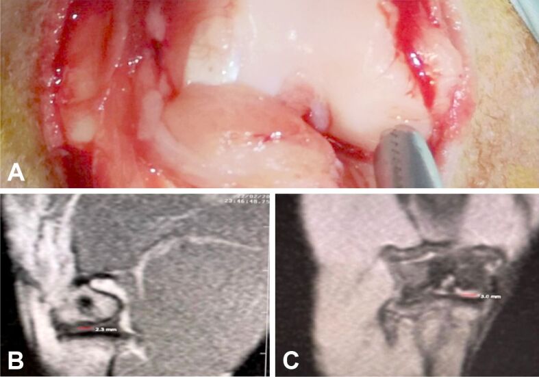
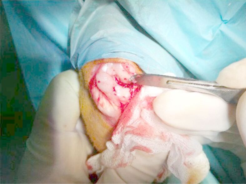
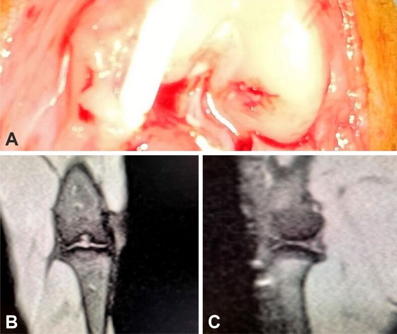
The present research study aimed to assess magnetic resonance imaging (MRI) changes and histological findings in the therapeutic effects of microfractures in the treatment of complex animal knee lesions resulting from osteochondral and meniscal defects resulting from non-total meniscectomies. The anterior cruciate ligament lesions are also proven to facilitate the development of osteoarthritis in the knee and worsen the prognosis. Surgery was performed on the right knee joint of 22 male rabbits in order to partially remove the anterior horn of the internal meniscus and to induce an osteochondral defect at the level of the internal femoral condyle. The induced lesion complex was aimed to simulate a clinical situation that occurs frequently in orthopedic practice when young adults undergo partial meniscectomy and at the time of surgery, an osteochondral defect is diagnosed. Rabbits were separated into two study groups: the control (C1) group and the microfractures (MF2) group. After the induced cartilage defect and partial meniscectomy, both groups were followed-up for six months using detailed MRI. Also, anatomical specimens were histologically analyzed to show modifications and signs of healing process, along with complications, in the study group. The results showed that the microfracture group had better results concerning articular surface defect healing in comparison to the control group. Our results suggest that microfractures do improve results concerning surface contact healing and serial MRI studies can be useful in observing the remodeling process in dynamics.
期刊介绍:
Romanian Journal of Morphology and Embryology (Rom J Morphol Embryol) publishes studies on all aspects of normal morphology and human comparative and experimental pathology. The Journal accepts only researches that utilize modern investigation methods (studies of anatomy, pathology, cytopathology, immunohistochemistry, histochemistry, immunology, morphometry, molecular and cellular biology, electronic microscopy, etc.).
 求助内容:
求助内容: 应助结果提醒方式:


