{"title":"利用 EPR 和 UV-vis 光谱以及氧化还原滴定法分析小漆酶中的电子传递途径","authors":"","doi":"10.1016/j.mrl.2024.200116","DOIUrl":null,"url":null,"abstract":"<div><p>Bacterial small laccases (SLAC) are promising industrial biocatalysts due to their ability to oxidize a broad range of substrates with exceptional thermostability and tolerance for alkaline pH. Electron transfer between substrate, copper centers, and O<sub>2</sub> is one of the key steps in the catalytic turnover of SLAC. However, limited research has been conducted on the electron transfer pathway of SLAC and SLAC-catalyzed reactions, hindering further engineering of SLAC to produce tunable biocatalysts for novel applications. Herein, the combinational use of electron paramagnetic resonance (EPR) and ultraviolet–visible (UV–vis) spectroscopic methods coupled with redox titration were employed to monitor the electron transfer processes and obtain further insights into the electron transfer pathway in SLAC. The reduction potentials for type 1 copper (T1Cu), type 2 copper (T2Cu) and type 3 copper (T3Cu) were determined to be 367 ± 2 mV, 378 ± 5 mV and 403 ± 2 mV, respectively. Moreover, the reduction potential of a selected substrate of SLAC, hydroquinone (HQ), was determined to be 288 mV using cyclic voltammetry (CV). In this way, an electron transfer pathway was identified based on the reduction potentials. Specifically, electrons are transferred from HQ to T1Cu, then to T2Cu and T3Cu, and finally to O<sub>2</sub>. Furthermore, superhyperfine splitting observed via EPR during redox titration indicated a modification in the covalency of T2Cu upon electron uptake, suggesting a conformational alteration in the protein environment surrounding the copper sites, which could potentially influence the reduction potential of the copper sites during catalytic processes. The results presented here not only provide a comprehensive method for analyzing the electron transfer pathway in metalloenzymes through reduction potential measurements, but also offer valuable insights for further engineering and directed evolution studies of SLAC in the aim for biotechnological and industrial applications.</p></div>","PeriodicalId":93594,"journal":{"name":"Magnetic Resonance Letters","volume":"4 3","pages":"Article 200116"},"PeriodicalIF":0.0000,"publicationDate":"2024-08-01","publicationTypes":"Journal Article","fieldsOfStudy":null,"isOpenAccess":false,"openAccessPdf":"https://www.sciencedirect.com/science/article/pii/S2772516224000238/pdfft?md5=9be3abce237719dfdcdc2795a0079a5f&pid=1-s2.0-S2772516224000238-main.pdf","citationCount":"0","resultStr":"{\"title\":\"Analysis of the electron transfer pathway in small laccase by EPR and UV–vis spectroscopy coupled with redox titration\",\"authors\":\"\",\"doi\":\"10.1016/j.mrl.2024.200116\",\"DOIUrl\":null,\"url\":null,\"abstract\":\"<div><p>Bacterial small laccases (SLAC) are promising industrial biocatalysts due to their ability to oxidize a broad range of substrates with exceptional thermostability and tolerance for alkaline pH. Electron transfer between substrate, copper centers, and O<sub>2</sub> is one of the key steps in the catalytic turnover of SLAC. However, limited research has been conducted on the electron transfer pathway of SLAC and SLAC-catalyzed reactions, hindering further engineering of SLAC to produce tunable biocatalysts for novel applications. Herein, the combinational use of electron paramagnetic resonance (EPR) and ultraviolet–visible (UV–vis) spectroscopic methods coupled with redox titration were employed to monitor the electron transfer processes and obtain further insights into the electron transfer pathway in SLAC. The reduction potentials for type 1 copper (T1Cu), type 2 copper (T2Cu) and type 3 copper (T3Cu) were determined to be 367 ± 2 mV, 378 ± 5 mV and 403 ± 2 mV, respectively. Moreover, the reduction potential of a selected substrate of SLAC, hydroquinone (HQ), was determined to be 288 mV using cyclic voltammetry (CV). In this way, an electron transfer pathway was identified based on the reduction potentials. Specifically, electrons are transferred from HQ to T1Cu, then to T2Cu and T3Cu, and finally to O<sub>2</sub>. Furthermore, superhyperfine splitting observed via EPR during redox titration indicated a modification in the covalency of T2Cu upon electron uptake, suggesting a conformational alteration in the protein environment surrounding the copper sites, which could potentially influence the reduction potential of the copper sites during catalytic processes. The results presented here not only provide a comprehensive method for analyzing the electron transfer pathway in metalloenzymes through reduction potential measurements, but also offer valuable insights for further engineering and directed evolution studies of SLAC in the aim for biotechnological and industrial applications.</p></div>\",\"PeriodicalId\":93594,\"journal\":{\"name\":\"Magnetic Resonance Letters\",\"volume\":\"4 3\",\"pages\":\"Article 200116\"},\"PeriodicalIF\":0.0000,\"publicationDate\":\"2024-08-01\",\"publicationTypes\":\"Journal Article\",\"fieldsOfStudy\":null,\"isOpenAccess\":false,\"openAccessPdf\":\"https://www.sciencedirect.com/science/article/pii/S2772516224000238/pdfft?md5=9be3abce237719dfdcdc2795a0079a5f&pid=1-s2.0-S2772516224000238-main.pdf\",\"citationCount\":\"0\",\"resultStr\":null,\"platform\":\"Semanticscholar\",\"paperid\":null,\"PeriodicalName\":\"Magnetic Resonance Letters\",\"FirstCategoryId\":\"1085\",\"ListUrlMain\":\"https://www.sciencedirect.com/science/article/pii/S2772516224000238\",\"RegionNum\":0,\"RegionCategory\":null,\"ArticlePicture\":[],\"TitleCN\":null,\"AbstractTextCN\":null,\"PMCID\":null,\"EPubDate\":\"\",\"PubModel\":\"\",\"JCR\":\"\",\"JCRName\":\"\",\"Score\":null,\"Total\":0}","platform":"Semanticscholar","paperid":null,"PeriodicalName":"Magnetic Resonance Letters","FirstCategoryId":"1085","ListUrlMain":"https://www.sciencedirect.com/science/article/pii/S2772516224000238","RegionNum":0,"RegionCategory":null,"ArticlePicture":[],"TitleCN":null,"AbstractTextCN":null,"PMCID":null,"EPubDate":"","PubModel":"","JCR":"","JCRName":"","Score":null,"Total":0}
Analysis of the electron transfer pathway in small laccase by EPR and UV–vis spectroscopy coupled with redox titration
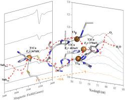
Bacterial small laccases (SLAC) are promising industrial biocatalysts due to their ability to oxidize a broad range of substrates with exceptional thermostability and tolerance for alkaline pH. Electron transfer between substrate, copper centers, and O2 is one of the key steps in the catalytic turnover of SLAC. However, limited research has been conducted on the electron transfer pathway of SLAC and SLAC-catalyzed reactions, hindering further engineering of SLAC to produce tunable biocatalysts for novel applications. Herein, the combinational use of electron paramagnetic resonance (EPR) and ultraviolet–visible (UV–vis) spectroscopic methods coupled with redox titration were employed to monitor the electron transfer processes and obtain further insights into the electron transfer pathway in SLAC. The reduction potentials for type 1 copper (T1Cu), type 2 copper (T2Cu) and type 3 copper (T3Cu) were determined to be 367 ± 2 mV, 378 ± 5 mV and 403 ± 2 mV, respectively. Moreover, the reduction potential of a selected substrate of SLAC, hydroquinone (HQ), was determined to be 288 mV using cyclic voltammetry (CV). In this way, an electron transfer pathway was identified based on the reduction potentials. Specifically, electrons are transferred from HQ to T1Cu, then to T2Cu and T3Cu, and finally to O2. Furthermore, superhyperfine splitting observed via EPR during redox titration indicated a modification in the covalency of T2Cu upon electron uptake, suggesting a conformational alteration in the protein environment surrounding the copper sites, which could potentially influence the reduction potential of the copper sites during catalytic processes. The results presented here not only provide a comprehensive method for analyzing the electron transfer pathway in metalloenzymes through reduction potential measurements, but also offer valuable insights for further engineering and directed evolution studies of SLAC in the aim for biotechnological and industrial applications.

 求助内容:
求助内容: 应助结果提醒方式:


