Yong-Hwan Cho, Jaehyung Choi, Chae-Wook Huh, Chang Hyeun Kim, Chul Hoon Chang, Soon Chan Kwon, Young Woo Kim, Seung Hun Sheen, Sukh Que Park, Jun Kyeung Ko, Sung-Kon Ha, Hae Woong Jeong, Hyen Seung Kang
{"title":"颅内动脉瘤血管内治疗后的影像随访策略:文献综述和指南建议。","authors":"Yong-Hwan Cho, Jaehyung Choi, Chae-Wook Huh, Chang Hyeun Kim, Chul Hoon Chang, Soon Chan Kwon, Young Woo Kim, Seung Hun Sheen, Sukh Que Park, Jun Kyeung Ko, Sung-Kon Ha, Hae Woong Jeong, Hyen Seung Kang","doi":"10.7461/jcen.2024.E2023.08.008","DOIUrl":null,"url":null,"abstract":"<p><strong>Objective: </strong>Endovascular coil embolization is the primary treatment modality for intracranial aneurysms. However, its long-term durability remains of concern, with a considerable proportion of cases requiring aneurysm reopening and retreatment. Therefore, establishing optimal follow-up imaging protocols is necessary to ensure a durable occlusion. This study aimed to develop guidelines for follow-up imaging strategies after endovascular treatment of intracranial aneurysms.</p><p><strong>Methods: </strong>A committee comprising members of the Korean Neuroendovascular Society and other relevant societies was formed. A literature review and analyses of the major published guidelines were conducted to gather evidence. A panel of 40 experts convened to achieve a consensus on the recommendations using the modified Delphi method.</p><p><strong>Results: </strong>The panel members reached the following consensus: 1. Schedule the initial follow-up imaging within 3-6 months of treatment. 2. Noninvasive imaging modalities, such as three-dimensional time-of-flight magnetic resonance angiography (MRA) or contrast-enhanced MRA, are alternatives to digital subtraction angiography (DSA) during the first follow-up. 3. Schedule mid-term follow-up imaging at 1, 2, 4, and 6 years after the initial treatment. 4. If noninvasive imaging reveals unstable changes in the treated aneurysms, DSA should be considered. 5. Consider late-term follow-up imaging every 3-5 years for lifelong monitoring of patients with unstable changes or at high risk of recurrence.</p><p><strong>Conclusions: </strong>The guidelines aim to provide physicians with the information to make informed decisions and provide patients with high-quality care. However, owing to a lack of specific recommendations and scientific data, these guidelines are based on expert consensus and should be considered in conjunction with individual patient characteristics and circumstances.</p>","PeriodicalId":94072,"journal":{"name":"Journal of cerebrovascular and endovascular neurosurgery","volume":" ","pages":"1-10"},"PeriodicalIF":0.0000,"publicationDate":"2024-03-01","publicationTypes":"Journal Article","fieldsOfStudy":null,"isOpenAccess":false,"openAccessPdf":"https://www.ncbi.nlm.nih.gov/pmc/articles/PMC10995472/pdf/","citationCount":"0","resultStr":"{\"title\":\"Imaging follow-up strategy after endovascular treatment of Intracranial aneurysms: A literature review and guideline recommendations.\",\"authors\":\"Yong-Hwan Cho, Jaehyung Choi, Chae-Wook Huh, Chang Hyeun Kim, Chul Hoon Chang, Soon Chan Kwon, Young Woo Kim, Seung Hun Sheen, Sukh Que Park, Jun Kyeung Ko, Sung-Kon Ha, Hae Woong Jeong, Hyen Seung Kang\",\"doi\":\"10.7461/jcen.2024.E2023.08.008\",\"DOIUrl\":null,\"url\":null,\"abstract\":\"<p><strong>Objective: </strong>Endovascular coil embolization is the primary treatment modality for intracranial aneurysms. However, its long-term durability remains of concern, with a considerable proportion of cases requiring aneurysm reopening and retreatment. Therefore, establishing optimal follow-up imaging protocols is necessary to ensure a durable occlusion. This study aimed to develop guidelines for follow-up imaging strategies after endovascular treatment of intracranial aneurysms.</p><p><strong>Methods: </strong>A committee comprising members of the Korean Neuroendovascular Society and other relevant societies was formed. A literature review and analyses of the major published guidelines were conducted to gather evidence. A panel of 40 experts convened to achieve a consensus on the recommendations using the modified Delphi method.</p><p><strong>Results: </strong>The panel members reached the following consensus: 1. Schedule the initial follow-up imaging within 3-6 months of treatment. 2. Noninvasive imaging modalities, such as three-dimensional time-of-flight magnetic resonance angiography (MRA) or contrast-enhanced MRA, are alternatives to digital subtraction angiography (DSA) during the first follow-up. 3. Schedule mid-term follow-up imaging at 1, 2, 4, and 6 years after the initial treatment. 4. If noninvasive imaging reveals unstable changes in the treated aneurysms, DSA should be considered. 5. Consider late-term follow-up imaging every 3-5 years for lifelong monitoring of patients with unstable changes or at high risk of recurrence.</p><p><strong>Conclusions: </strong>The guidelines aim to provide physicians with the information to make informed decisions and provide patients with high-quality care. However, owing to a lack of specific recommendations and scientific data, these guidelines are based on expert consensus and should be considered in conjunction with individual patient characteristics and circumstances.</p>\",\"PeriodicalId\":94072,\"journal\":{\"name\":\"Journal of cerebrovascular and endovascular neurosurgery\",\"volume\":\" \",\"pages\":\"1-10\"},\"PeriodicalIF\":0.0000,\"publicationDate\":\"2024-03-01\",\"publicationTypes\":\"Journal Article\",\"fieldsOfStudy\":null,\"isOpenAccess\":false,\"openAccessPdf\":\"https://www.ncbi.nlm.nih.gov/pmc/articles/PMC10995472/pdf/\",\"citationCount\":\"0\",\"resultStr\":null,\"platform\":\"Semanticscholar\",\"paperid\":null,\"PeriodicalName\":\"Journal of cerebrovascular and endovascular neurosurgery\",\"FirstCategoryId\":\"1085\",\"ListUrlMain\":\"https://doi.org/10.7461/jcen.2024.E2023.08.008\",\"RegionNum\":0,\"RegionCategory\":null,\"ArticlePicture\":[],\"TitleCN\":null,\"AbstractTextCN\":null,\"PMCID\":null,\"EPubDate\":\"2024/3/25 0:00:00\",\"PubModel\":\"Epub\",\"JCR\":\"\",\"JCRName\":\"\",\"Score\":null,\"Total\":0}","platform":"Semanticscholar","paperid":null,"PeriodicalName":"Journal of cerebrovascular and endovascular neurosurgery","FirstCategoryId":"1085","ListUrlMain":"https://doi.org/10.7461/jcen.2024.E2023.08.008","RegionNum":0,"RegionCategory":null,"ArticlePicture":[],"TitleCN":null,"AbstractTextCN":null,"PMCID":null,"EPubDate":"2024/3/25 0:00:00","PubModel":"Epub","JCR":"","JCRName":"","Score":null,"Total":0}
Imaging follow-up strategy after endovascular treatment of Intracranial aneurysms: A literature review and guideline recommendations.
Objective: Endovascular coil embolization is the primary treatment modality for intracranial aneurysms. However, its long-term durability remains of concern, with a considerable proportion of cases requiring aneurysm reopening and retreatment. Therefore, establishing optimal follow-up imaging protocols is necessary to ensure a durable occlusion. This study aimed to develop guidelines for follow-up imaging strategies after endovascular treatment of intracranial aneurysms.
Methods: A committee comprising members of the Korean Neuroendovascular Society and other relevant societies was formed. A literature review and analyses of the major published guidelines were conducted to gather evidence. A panel of 40 experts convened to achieve a consensus on the recommendations using the modified Delphi method.
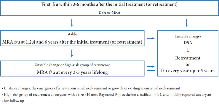
Results: The panel members reached the following consensus: 1. Schedule the initial follow-up imaging within 3-6 months of treatment. 2. Noninvasive imaging modalities, such as three-dimensional time-of-flight magnetic resonance angiography (MRA) or contrast-enhanced MRA, are alternatives to digital subtraction angiography (DSA) during the first follow-up. 3. Schedule mid-term follow-up imaging at 1, 2, 4, and 6 years after the initial treatment. 4. If noninvasive imaging reveals unstable changes in the treated aneurysms, DSA should be considered. 5. Consider late-term follow-up imaging every 3-5 years for lifelong monitoring of patients with unstable changes or at high risk of recurrence.
Conclusions: The guidelines aim to provide physicians with the information to make informed decisions and provide patients with high-quality care. However, owing to a lack of specific recommendations and scientific data, these guidelines are based on expert consensus and should be considered in conjunction with individual patient characteristics and circumstances.

 求助内容:
求助内容: 应助结果提醒方式:


