{"title":"当深度学习还不够时:人工生命作为乳腺癌超声波图像分割的辅助工具。","authors":"Nalan Karunanayake, Stanislav S Makhanov","doi":"10.1007/s11517-024-03026-x","DOIUrl":null,"url":null,"abstract":"<p><p>Segmentation of tumors in ultrasound (US) images of the breast is a critical issue in medical imaging. Due to the poor quality of US images and the varying specifications of US machines, segmentation and classification of abnormalities present difficulties even for trained radiologists. The paper aims to introduce a novel AI-based hybrid model for US segmentation that offers high accuracy, requires relatively smaller datasets, and is capable of handling previously unseen data. The software can be used for diagnostics and the US-guided biopsies. A unique and robust hybrid approach that combines deep learning (DL) and multi-agent artificial life (AL) has been introduced. The algorithms are verified on three US datasets. The method outperforms 14 selected state-of-the-art algorithms applied to US images characterized by complex geometry and high level of noise. The paper offers an original classification of the images and tests to analyze the limits of the DL. The model has been trained and verified on 1264 ultrasound images. The images are in the JPEG and PNG formats. The age of the patients ranges from 22 to 73 years. The 14 benchmark algorithms include deformable shapes, edge linking, superpixels, machine learning, and DL methods. The tests use eight-region shape- and contour-based evaluation metrics. The proposed method (DL-AL) produces excellent results in terms of the dice coefficient (region) and the relative Hausdorff distance H<sub>3</sub> (contour-based) as follows: the easiest image complexity level, Dice = 0.96 and H<sub>3</sub> = 0.26; the medium complexity level, Dice = 0.91 and H<sub>3</sub> = 0.82; and the hardest complexity level, Dice = 0.90 and H<sub>3</sub> = 0.84. All other metrics follow the same pattern. The DL-AL outperforms the second best (Unet-based) method by 10-20%. The method has been also tested by a series of unconventional tests. The model was trained on low complexity images and applied to the entire set of images. These results are summarized below. (1) Only the low complexity images have been used for training (68% unknown images): Dice = 0.80 and H<sub>3</sub> = 2.01. (2) The low and the medium complexity images have been used for training (51% unknown images): Dice = 0.86 and H<sub>3</sub> = 1.32. (3) The low, medium, and hard complexity images have been used for training (35% unknown images): Dice = 0.92 and H<sub>3</sub> = 0.76. These tests show a significant advantage of DL-AL over 30%. A video demo illustrating the algorithm is at http://tinyurl.com/mr4ah687 .</p>","PeriodicalId":49840,"journal":{"name":"Medical & Biological Engineering & Computing","volume":" ","pages":"2497-2520"},"PeriodicalIF":2.6000,"publicationDate":"2025-08-01","publicationTypes":"Journal Article","fieldsOfStudy":null,"isOpenAccess":false,"openAccessPdf":"","citationCount":"0","resultStr":"{\"title\":\"When deep learning is not enough: artificial life as a supplementary tool for segmentation of ultrasound images of breast cancer.\",\"authors\":\"Nalan Karunanayake, Stanislav S Makhanov\",\"doi\":\"10.1007/s11517-024-03026-x\",\"DOIUrl\":null,\"url\":null,\"abstract\":\"<p><p>Segmentation of tumors in ultrasound (US) images of the breast is a critical issue in medical imaging. Due to the poor quality of US images and the varying specifications of US machines, segmentation and classification of abnormalities present difficulties even for trained radiologists. The paper aims to introduce a novel AI-based hybrid model for US segmentation that offers high accuracy, requires relatively smaller datasets, and is capable of handling previously unseen data. The software can be used for diagnostics and the US-guided biopsies. A unique and robust hybrid approach that combines deep learning (DL) and multi-agent artificial life (AL) has been introduced. The algorithms are verified on three US datasets. The method outperforms 14 selected state-of-the-art algorithms applied to US images characterized by complex geometry and high level of noise. The paper offers an original classification of the images and tests to analyze the limits of the DL. The model has been trained and verified on 1264 ultrasound images. The images are in the JPEG and PNG formats. The age of the patients ranges from 22 to 73 years. The 14 benchmark algorithms include deformable shapes, edge linking, superpixels, machine learning, and DL methods. The tests use eight-region shape- and contour-based evaluation metrics. The proposed method (DL-AL) produces excellent results in terms of the dice coefficient (region) and the relative Hausdorff distance H<sub>3</sub> (contour-based) as follows: the easiest image complexity level, Dice = 0.96 and H<sub>3</sub> = 0.26; the medium complexity level, Dice = 0.91 and H<sub>3</sub> = 0.82; and the hardest complexity level, Dice = 0.90 and H<sub>3</sub> = 0.84. All other metrics follow the same pattern. The DL-AL outperforms the second best (Unet-based) method by 10-20%. The method has been also tested by a series of unconventional tests. The model was trained on low complexity images and applied to the entire set of images. These results are summarized below. (1) Only the low complexity images have been used for training (68% unknown images): Dice = 0.80 and H<sub>3</sub> = 2.01. (2) The low and the medium complexity images have been used for training (51% unknown images): Dice = 0.86 and H<sub>3</sub> = 1.32. (3) The low, medium, and hard complexity images have been used for training (35% unknown images): Dice = 0.92 and H<sub>3</sub> = 0.76. These tests show a significant advantage of DL-AL over 30%. A video demo illustrating the algorithm is at http://tinyurl.com/mr4ah687 .</p>\",\"PeriodicalId\":49840,\"journal\":{\"name\":\"Medical & Biological Engineering & Computing\",\"volume\":\" \",\"pages\":\"2497-2520\"},\"PeriodicalIF\":2.6000,\"publicationDate\":\"2025-08-01\",\"publicationTypes\":\"Journal Article\",\"fieldsOfStudy\":null,\"isOpenAccess\":false,\"openAccessPdf\":\"\",\"citationCount\":\"0\",\"resultStr\":null,\"platform\":\"Semanticscholar\",\"paperid\":null,\"PeriodicalName\":\"Medical & Biological Engineering & Computing\",\"FirstCategoryId\":\"5\",\"ListUrlMain\":\"https://doi.org/10.1007/s11517-024-03026-x\",\"RegionNum\":4,\"RegionCategory\":\"医学\",\"ArticlePicture\":[],\"TitleCN\":null,\"AbstractTextCN\":null,\"PMCID\":null,\"EPubDate\":\"2024/3/18 0:00:00\",\"PubModel\":\"Epub\",\"JCR\":\"Q2\",\"JCRName\":\"COMPUTER SCIENCE, INTERDISCIPLINARY APPLICATIONS\",\"Score\":null,\"Total\":0}","platform":"Semanticscholar","paperid":null,"PeriodicalName":"Medical & Biological Engineering & Computing","FirstCategoryId":"5","ListUrlMain":"https://doi.org/10.1007/s11517-024-03026-x","RegionNum":4,"RegionCategory":"医学","ArticlePicture":[],"TitleCN":null,"AbstractTextCN":null,"PMCID":null,"EPubDate":"2024/3/18 0:00:00","PubModel":"Epub","JCR":"Q2","JCRName":"COMPUTER SCIENCE, INTERDISCIPLINARY APPLICATIONS","Score":null,"Total":0}
When deep learning is not enough: artificial life as a supplementary tool for segmentation of ultrasound images of breast cancer.
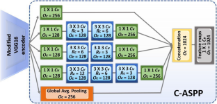
Segmentation of tumors in ultrasound (US) images of the breast is a critical issue in medical imaging. Due to the poor quality of US images and the varying specifications of US machines, segmentation and classification of abnormalities present difficulties even for trained radiologists. The paper aims to introduce a novel AI-based hybrid model for US segmentation that offers high accuracy, requires relatively smaller datasets, and is capable of handling previously unseen data. The software can be used for diagnostics and the US-guided biopsies. A unique and robust hybrid approach that combines deep learning (DL) and multi-agent artificial life (AL) has been introduced. The algorithms are verified on three US datasets. The method outperforms 14 selected state-of-the-art algorithms applied to US images characterized by complex geometry and high level of noise. The paper offers an original classification of the images and tests to analyze the limits of the DL. The model has been trained and verified on 1264 ultrasound images. The images are in the JPEG and PNG formats. The age of the patients ranges from 22 to 73 years. The 14 benchmark algorithms include deformable shapes, edge linking, superpixels, machine learning, and DL methods. The tests use eight-region shape- and contour-based evaluation metrics. The proposed method (DL-AL) produces excellent results in terms of the dice coefficient (region) and the relative Hausdorff distance H3 (contour-based) as follows: the easiest image complexity level, Dice = 0.96 and H3 = 0.26; the medium complexity level, Dice = 0.91 and H3 = 0.82; and the hardest complexity level, Dice = 0.90 and H3 = 0.84. All other metrics follow the same pattern. The DL-AL outperforms the second best (Unet-based) method by 10-20%. The method has been also tested by a series of unconventional tests. The model was trained on low complexity images and applied to the entire set of images. These results are summarized below. (1) Only the low complexity images have been used for training (68% unknown images): Dice = 0.80 and H3 = 2.01. (2) The low and the medium complexity images have been used for training (51% unknown images): Dice = 0.86 and H3 = 1.32. (3) The low, medium, and hard complexity images have been used for training (35% unknown images): Dice = 0.92 and H3 = 0.76. These tests show a significant advantage of DL-AL over 30%. A video demo illustrating the algorithm is at http://tinyurl.com/mr4ah687 .
期刊介绍:
Founded in 1963, Medical & Biological Engineering & Computing (MBEC) continues to serve the biomedical engineering community, covering the entire spectrum of biomedical and clinical engineering. The journal presents exciting and vital experimental and theoretical developments in biomedical science and technology, and reports on advances in computer-based methodologies in these multidisciplinary subjects. The journal also incorporates new and evolving technologies including cellular engineering and molecular imaging.
MBEC publishes original research articles as well as reviews and technical notes. Its Rapid Communications category focuses on material of immediate value to the readership, while the Controversies section provides a forum to exchange views on selected issues, stimulating a vigorous and informed debate in this exciting and high profile field.
MBEC is an official journal of the International Federation of Medical and Biological Engineering (IFMBE).

 求助内容:
求助内容: 应助结果提醒方式:


