Cyan Ching, Julien Maufront, Aurélie di Cicco, Daniel Lévy, Manuela Dezi
{"title":"冷接触:膜接触点及其组件的冷冻电子显微镜观察","authors":"Cyan Ching, Julien Maufront, Aurélie di Cicco, Daniel Lévy, Manuela Dezi","doi":"10.1177/25152564241231364","DOIUrl":null,"url":null,"abstract":"<p><p>Electron microscopy has played a pivotal role in elucidating the ultrastructure of membrane contact sites between cellular organelles. The advent of cryo-electron microscopy has ushered in the ability to determine atomic models of constituent proteins or protein complexes within sites of membrane contact through single particle analysis. Furthermore, it enables the visualization of the three-dimensional architecture of membrane contact sites, encompassing numerous copies of proteins, whether in vitro reconstituted or directly observed in situ using cryo-electron tomography. Nevertheless, there exists a scarcity of cryo-electron microscopy studies focused on the site of membrane contact and their constitutive proteins. This review provides an overview of the contributions made by cryo-electron microscopy to our understanding of membrane contact sites, outlines the associated limitations, and explores prospects in this field.</p>","PeriodicalId":101304,"journal":{"name":"Contact (Thousand Oaks (Ventura County, Calif.))","volume":"7 ","pages":"25152564241231364"},"PeriodicalIF":0.0000,"publicationDate":"2024-02-25","publicationTypes":"Journal Article","fieldsOfStudy":null,"isOpenAccess":false,"openAccessPdf":"https://www.ncbi.nlm.nih.gov/pmc/articles/PMC10895918/pdf/","citationCount":"0","resultStr":"{\"title\":\"C<i>ool-contacts</i>: Cryo-Electron Microscopy of Membrane Contact Sites and Their Components.\",\"authors\":\"Cyan Ching, Julien Maufront, Aurélie di Cicco, Daniel Lévy, Manuela Dezi\",\"doi\":\"10.1177/25152564241231364\",\"DOIUrl\":null,\"url\":null,\"abstract\":\"<p><p>Electron microscopy has played a pivotal role in elucidating the ultrastructure of membrane contact sites between cellular organelles. The advent of cryo-electron microscopy has ushered in the ability to determine atomic models of constituent proteins or protein complexes within sites of membrane contact through single particle analysis. Furthermore, it enables the visualization of the three-dimensional architecture of membrane contact sites, encompassing numerous copies of proteins, whether in vitro reconstituted or directly observed in situ using cryo-electron tomography. Nevertheless, there exists a scarcity of cryo-electron microscopy studies focused on the site of membrane contact and their constitutive proteins. This review provides an overview of the contributions made by cryo-electron microscopy to our understanding of membrane contact sites, outlines the associated limitations, and explores prospects in this field.</p>\",\"PeriodicalId\":101304,\"journal\":{\"name\":\"Contact (Thousand Oaks (Ventura County, Calif.))\",\"volume\":\"7 \",\"pages\":\"25152564241231364\"},\"PeriodicalIF\":0.0000,\"publicationDate\":\"2024-02-25\",\"publicationTypes\":\"Journal Article\",\"fieldsOfStudy\":null,\"isOpenAccess\":false,\"openAccessPdf\":\"https://www.ncbi.nlm.nih.gov/pmc/articles/PMC10895918/pdf/\",\"citationCount\":\"0\",\"resultStr\":null,\"platform\":\"Semanticscholar\",\"paperid\":null,\"PeriodicalName\":\"Contact (Thousand Oaks (Ventura County, Calif.))\",\"FirstCategoryId\":\"1085\",\"ListUrlMain\":\"https://doi.org/10.1177/25152564241231364\",\"RegionNum\":0,\"RegionCategory\":null,\"ArticlePicture\":[],\"TitleCN\":null,\"AbstractTextCN\":null,\"PMCID\":null,\"EPubDate\":\"2024/1/1 0:00:00\",\"PubModel\":\"eCollection\",\"JCR\":\"\",\"JCRName\":\"\",\"Score\":null,\"Total\":0}","platform":"Semanticscholar","paperid":null,"PeriodicalName":"Contact (Thousand Oaks (Ventura County, Calif.))","FirstCategoryId":"1085","ListUrlMain":"https://doi.org/10.1177/25152564241231364","RegionNum":0,"RegionCategory":null,"ArticlePicture":[],"TitleCN":null,"AbstractTextCN":null,"PMCID":null,"EPubDate":"2024/1/1 0:00:00","PubModel":"eCollection","JCR":"","JCRName":"","Score":null,"Total":0}
Cool-contacts: Cryo-Electron Microscopy of Membrane Contact Sites and Their Components.
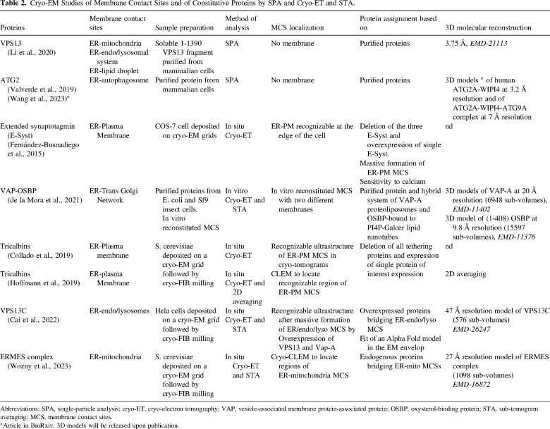
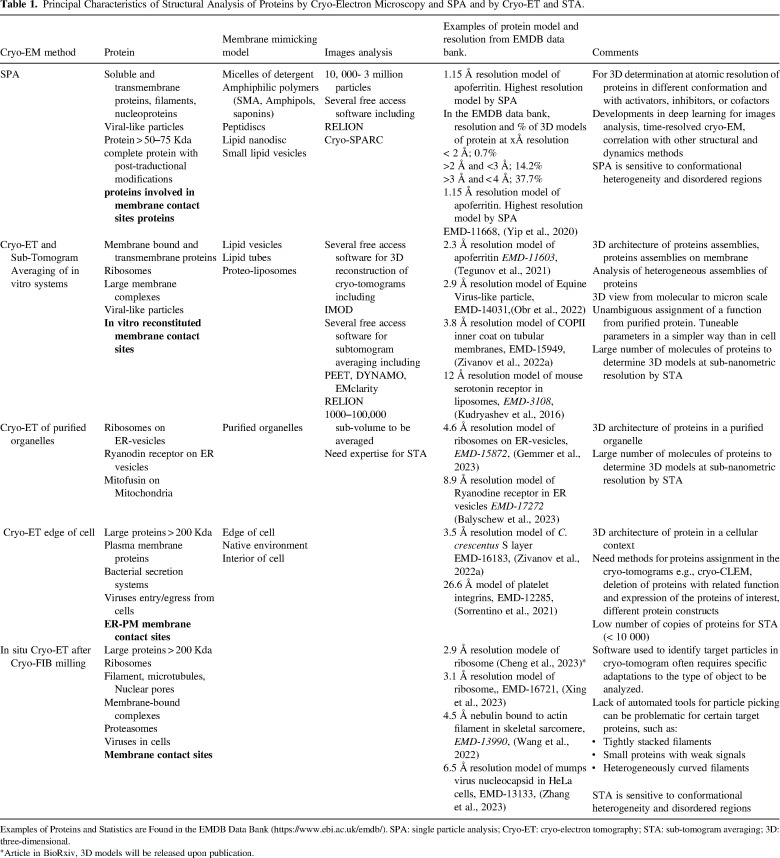
Electron microscopy has played a pivotal role in elucidating the ultrastructure of membrane contact sites between cellular organelles. The advent of cryo-electron microscopy has ushered in the ability to determine atomic models of constituent proteins or protein complexes within sites of membrane contact through single particle analysis. Furthermore, it enables the visualization of the three-dimensional architecture of membrane contact sites, encompassing numerous copies of proteins, whether in vitro reconstituted or directly observed in situ using cryo-electron tomography. Nevertheless, there exists a scarcity of cryo-electron microscopy studies focused on the site of membrane contact and their constitutive proteins. This review provides an overview of the contributions made by cryo-electron microscopy to our understanding of membrane contact sites, outlines the associated limitations, and explores prospects in this field.

 求助内容:
求助内容: 应助结果提醒方式:


