Zeynep Özbek, Banu Lebe, Mustafa Kayabaşı, Ali Osman Saatci
{"title":"模仿结节性前巩膜炎的嵌入式巩膜异物","authors":"Zeynep Özbek, Banu Lebe, Mustafa Kayabaşı, Ali Osman Saatci","doi":"10.4274/tjo.galenos.2023.37460","DOIUrl":null,"url":null,"abstract":"<p><p>A 56-year-old man was referred to our clinic for unilateral nodular scleritis unresponsive to systemic corticosteroids. A localized, nodular hyperemia on the nasal bulbar conjunctiva surrounding a central cyst-like lesion together with vascular engorgement was observed on slit-lamp examination of the left eye. No abnormal fundoscopic findings were noted. Surgical exploration revealed an embedded episcleral brown colored, soft to touch, splinter-like organic foreign body (FB) which was confirmed by the histopathological examination. Nodular hyperemia resolved during the postoperative follow-up period, and mild scar tissue accompanied by scleral thinning developed in the left nasal bulbar conjunctiva. Ocular injury associated with FBs may cause significant ocular morbidity depending on the nature and location of the FB. Severe visual disability may occur if left untreated. Subconjunctival FBs are rare and may present with a clinical picture mimicking episcleritis or scleritis. History of trauma involving a FB should always be assessed for an accurate differential diagnosis and appropriate management of patients with anterior scleritis.</p>","PeriodicalId":23373,"journal":{"name":"Turkish Journal of Ophthalmology","volume":"54 1","pages":"46-48"},"PeriodicalIF":0.0000,"publicationDate":"2024-02-22","publicationTypes":"Journal Article","fieldsOfStudy":null,"isOpenAccess":false,"openAccessPdf":"https://www.ncbi.nlm.nih.gov/pmc/articles/PMC10895169/pdf/","citationCount":"0","resultStr":"{\"title\":\"Embedded Episcleral Foreign Body Mimicking Nodular Anterior Scleritis.\",\"authors\":\"Zeynep Özbek, Banu Lebe, Mustafa Kayabaşı, Ali Osman Saatci\",\"doi\":\"10.4274/tjo.galenos.2023.37460\",\"DOIUrl\":null,\"url\":null,\"abstract\":\"<p><p>A 56-year-old man was referred to our clinic for unilateral nodular scleritis unresponsive to systemic corticosteroids. A localized, nodular hyperemia on the nasal bulbar conjunctiva surrounding a central cyst-like lesion together with vascular engorgement was observed on slit-lamp examination of the left eye. No abnormal fundoscopic findings were noted. Surgical exploration revealed an embedded episcleral brown colored, soft to touch, splinter-like organic foreign body (FB) which was confirmed by the histopathological examination. Nodular hyperemia resolved during the postoperative follow-up period, and mild scar tissue accompanied by scleral thinning developed in the left nasal bulbar conjunctiva. Ocular injury associated with FBs may cause significant ocular morbidity depending on the nature and location of the FB. Severe visual disability may occur if left untreated. Subconjunctival FBs are rare and may present with a clinical picture mimicking episcleritis or scleritis. History of trauma involving a FB should always be assessed for an accurate differential diagnosis and appropriate management of patients with anterior scleritis.</p>\",\"PeriodicalId\":23373,\"journal\":{\"name\":\"Turkish Journal of Ophthalmology\",\"volume\":\"54 1\",\"pages\":\"46-48\"},\"PeriodicalIF\":0.0000,\"publicationDate\":\"2024-02-22\",\"publicationTypes\":\"Journal Article\",\"fieldsOfStudy\":null,\"isOpenAccess\":false,\"openAccessPdf\":\"https://www.ncbi.nlm.nih.gov/pmc/articles/PMC10895169/pdf/\",\"citationCount\":\"0\",\"resultStr\":null,\"platform\":\"Semanticscholar\",\"paperid\":null,\"PeriodicalName\":\"Turkish Journal of Ophthalmology\",\"FirstCategoryId\":\"1085\",\"ListUrlMain\":\"https://doi.org/10.4274/tjo.galenos.2023.37460\",\"RegionNum\":0,\"RegionCategory\":null,\"ArticlePicture\":[],\"TitleCN\":null,\"AbstractTextCN\":null,\"PMCID\":null,\"EPubDate\":\"\",\"PubModel\":\"\",\"JCR\":\"Q3\",\"JCRName\":\"Medicine\",\"Score\":null,\"Total\":0}","platform":"Semanticscholar","paperid":null,"PeriodicalName":"Turkish Journal of Ophthalmology","FirstCategoryId":"1085","ListUrlMain":"https://doi.org/10.4274/tjo.galenos.2023.37460","RegionNum":0,"RegionCategory":null,"ArticlePicture":[],"TitleCN":null,"AbstractTextCN":null,"PMCID":null,"EPubDate":"","PubModel":"","JCR":"Q3","JCRName":"Medicine","Score":null,"Total":0}
Embedded Episcleral Foreign Body Mimicking Nodular Anterior Scleritis.
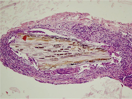
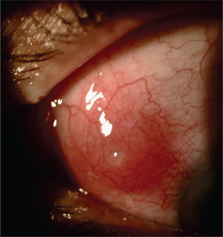
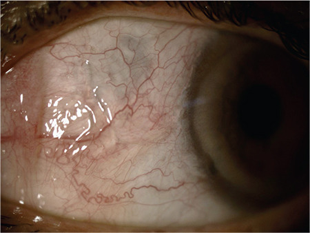
A 56-year-old man was referred to our clinic for unilateral nodular scleritis unresponsive to systemic corticosteroids. A localized, nodular hyperemia on the nasal bulbar conjunctiva surrounding a central cyst-like lesion together with vascular engorgement was observed on slit-lamp examination of the left eye. No abnormal fundoscopic findings were noted. Surgical exploration revealed an embedded episcleral brown colored, soft to touch, splinter-like organic foreign body (FB) which was confirmed by the histopathological examination. Nodular hyperemia resolved during the postoperative follow-up period, and mild scar tissue accompanied by scleral thinning developed in the left nasal bulbar conjunctiva. Ocular injury associated with FBs may cause significant ocular morbidity depending on the nature and location of the FB. Severe visual disability may occur if left untreated. Subconjunctival FBs are rare and may present with a clinical picture mimicking episcleritis or scleritis. History of trauma involving a FB should always be assessed for an accurate differential diagnosis and appropriate management of patients with anterior scleritis.
期刊介绍:
The Turkish Journal of Ophthalmology (TJO) is the only scientific periodical publication of the Turkish Ophthalmological Association and has been published since January 1929. In its early years, the journal was published in Turkish and French. Although there were temporary interruptions in the publication of the journal due to various challenges, the Turkish Journal of Ophthalmology has been published continually from 1971 to the present. The target audience includes specialists and physicians in training in ophthalmology in all relevant disciplines.

 求助内容:
求助内容: 应助结果提醒方式:


