Pae Sun Suh, So Yeong Jeong, Jung Hwan Baek, Tae Yong Kim, Yu-Mi Lee, Dong Eun Song, Yun Seo Park, Je Young Ahn, Sae Rom Chung, Young Jun Choi, Jeong Hyun Lee
{"title":"结耳征:甲状腺手术后缝合肉芽肿的超声诊断特征。","authors":"Pae Sun Suh, So Yeong Jeong, Jung Hwan Baek, Tae Yong Kim, Yu-Mi Lee, Dong Eun Song, Yun Seo Park, Je Young Ahn, Sae Rom Chung, Young Jun Choi, Jeong Hyun Lee","doi":"10.14366/usg.23210","DOIUrl":null,"url":null,"abstract":"<p><strong>Purpose: </strong>This study investigated the clinical and ultrasonographic (US) findings of suture granulomas and recurrent tumors, and aimed to identify specific characteristics of suture granulomas through an experimental study.</p><p><strong>Methods: </strong>This retrospective study included 20 pathologically confirmed suture granulomas and 40 recurrent tumors between January 2010 and December 2020. The clinical findings included suture material, surgery, and initial TNM stage. The US findings included shape, size, margin, echogenicity, heterogeneity, vascularity, and internal echogenic foci. The distribution, paired appearance, and \"knot-and-ear\" appearance of internal echogenic foci were assessed. An experiment using pork meat investigated the US configuration of suture knots.</p><p><strong>Results: </strong>Eighteen patients with 20 suture granulomas (15 women; mean age, 52±13 years) and 37 patients with 40 recurrent tumors (24 women; 54±18 years) were included. Patients with suture granulomas exhibited earlier initial T and N stages than those with recurrent tumors. The knot-and-ear appearance, defined as an echogenic dot accompanied by two adjacent echogenic dots or lines based on experimental findings, was observed in 50% of suture granulomas, but not in recurrent tumors (P<0.001). Central internal echogenic foci (68.8%, P=0.023) and paired appearance (75.0%, P<0.001) were more frequent in suture granulomas. During follow-up, 94.1% of suture granulomas shrunk.</p><p><strong>Conclusion: </strong>The knot-and-ear appearance is a potential pathognomonic finding of suture granuloma, and central internal echogenic foci with paired appearance were typical US features. Clinically, suture granulomas showed early T and N stages and size reduction during follow-up. Understanding these features can prevent unnecessary biopsy or diagnostic surgery.</p>","PeriodicalId":54227,"journal":{"name":"Ultrasonography","volume":" ","pages":"141-150"},"PeriodicalIF":2.5000,"publicationDate":"2024-03-01","publicationTypes":"Journal Article","fieldsOfStudy":null,"isOpenAccess":false,"openAccessPdf":"https://www.ncbi.nlm.nih.gov/pmc/articles/PMC10915117/pdf/","citationCount":"0","resultStr":"{\"title\":\"Knot-and-ear sign: a pathognomonic ultrasonographic feature of suture granuloma after thyroid surgery.\",\"authors\":\"Pae Sun Suh, So Yeong Jeong, Jung Hwan Baek, Tae Yong Kim, Yu-Mi Lee, Dong Eun Song, Yun Seo Park, Je Young Ahn, Sae Rom Chung, Young Jun Choi, Jeong Hyun Lee\",\"doi\":\"10.14366/usg.23210\",\"DOIUrl\":null,\"url\":null,\"abstract\":\"<p><strong>Purpose: </strong>This study investigated the clinical and ultrasonographic (US) findings of suture granulomas and recurrent tumors, and aimed to identify specific characteristics of suture granulomas through an experimental study.</p><p><strong>Methods: </strong>This retrospective study included 20 pathologically confirmed suture granulomas and 40 recurrent tumors between January 2010 and December 2020. The clinical findings included suture material, surgery, and initial TNM stage. The US findings included shape, size, margin, echogenicity, heterogeneity, vascularity, and internal echogenic foci. The distribution, paired appearance, and \\\"knot-and-ear\\\" appearance of internal echogenic foci were assessed. An experiment using pork meat investigated the US configuration of suture knots.</p><p><strong>Results: </strong>Eighteen patients with 20 suture granulomas (15 women; mean age, 52±13 years) and 37 patients with 40 recurrent tumors (24 women; 54±18 years) were included. Patients with suture granulomas exhibited earlier initial T and N stages than those with recurrent tumors. The knot-and-ear appearance, defined as an echogenic dot accompanied by two adjacent echogenic dots or lines based on experimental findings, was observed in 50% of suture granulomas, but not in recurrent tumors (P<0.001). Central internal echogenic foci (68.8%, P=0.023) and paired appearance (75.0%, P<0.001) were more frequent in suture granulomas. During follow-up, 94.1% of suture granulomas shrunk.</p><p><strong>Conclusion: </strong>The knot-and-ear appearance is a potential pathognomonic finding of suture granuloma, and central internal echogenic foci with paired appearance were typical US features. Clinically, suture granulomas showed early T and N stages and size reduction during follow-up. Understanding these features can prevent unnecessary biopsy or diagnostic surgery.</p>\",\"PeriodicalId\":54227,\"journal\":{\"name\":\"Ultrasonography\",\"volume\":\" \",\"pages\":\"141-150\"},\"PeriodicalIF\":2.5000,\"publicationDate\":\"2024-03-01\",\"publicationTypes\":\"Journal Article\",\"fieldsOfStudy\":null,\"isOpenAccess\":false,\"openAccessPdf\":\"https://www.ncbi.nlm.nih.gov/pmc/articles/PMC10915117/pdf/\",\"citationCount\":\"0\",\"resultStr\":null,\"platform\":\"Semanticscholar\",\"paperid\":null,\"PeriodicalName\":\"Ultrasonography\",\"FirstCategoryId\":\"3\",\"ListUrlMain\":\"https://doi.org/10.14366/usg.23210\",\"RegionNum\":3,\"RegionCategory\":\"医学\",\"ArticlePicture\":[],\"TitleCN\":null,\"AbstractTextCN\":null,\"PMCID\":null,\"EPubDate\":\"2024/1/23 0:00:00\",\"PubModel\":\"Epub\",\"JCR\":\"Q2\",\"JCRName\":\"RADIOLOGY, NUCLEAR MEDICINE & MEDICAL IMAGING\",\"Score\":null,\"Total\":0}","platform":"Semanticscholar","paperid":null,"PeriodicalName":"Ultrasonography","FirstCategoryId":"3","ListUrlMain":"https://doi.org/10.14366/usg.23210","RegionNum":3,"RegionCategory":"医学","ArticlePicture":[],"TitleCN":null,"AbstractTextCN":null,"PMCID":null,"EPubDate":"2024/1/23 0:00:00","PubModel":"Epub","JCR":"Q2","JCRName":"RADIOLOGY, NUCLEAR MEDICINE & MEDICAL IMAGING","Score":null,"Total":0}
Knot-and-ear sign: a pathognomonic ultrasonographic feature of suture granuloma after thyroid surgery.
Purpose: This study investigated the clinical and ultrasonographic (US) findings of suture granulomas and recurrent tumors, and aimed to identify specific characteristics of suture granulomas through an experimental study.
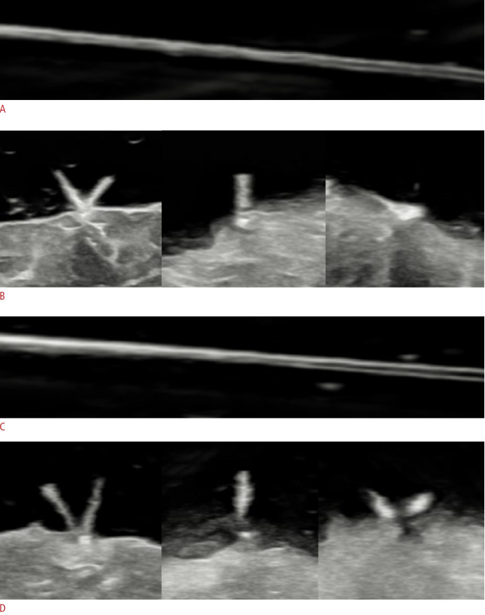
Methods: This retrospective study included 20 pathologically confirmed suture granulomas and 40 recurrent tumors between January 2010 and December 2020. The clinical findings included suture material, surgery, and initial TNM stage. The US findings included shape, size, margin, echogenicity, heterogeneity, vascularity, and internal echogenic foci. The distribution, paired appearance, and "knot-and-ear" appearance of internal echogenic foci were assessed. An experiment using pork meat investigated the US configuration of suture knots.
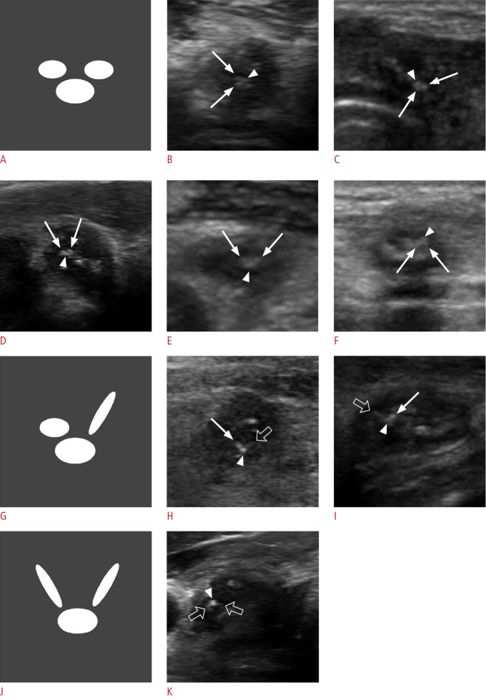
Results: Eighteen patients with 20 suture granulomas (15 women; mean age, 52±13 years) and 37 patients with 40 recurrent tumors (24 women; 54±18 years) were included. Patients with suture granulomas exhibited earlier initial T and N stages than those with recurrent tumors. The knot-and-ear appearance, defined as an echogenic dot accompanied by two adjacent echogenic dots or lines based on experimental findings, was observed in 50% of suture granulomas, but not in recurrent tumors (P<0.001). Central internal echogenic foci (68.8%, P=0.023) and paired appearance (75.0%, P<0.001) were more frequent in suture granulomas. During follow-up, 94.1% of suture granulomas shrunk.
Conclusion: The knot-and-ear appearance is a potential pathognomonic finding of suture granuloma, and central internal echogenic foci with paired appearance were typical US features. Clinically, suture granulomas showed early T and N stages and size reduction during follow-up. Understanding these features can prevent unnecessary biopsy or diagnostic surgery.
UltrasonographyMedicine-Radiology, Nuclear Medicine and Imaging
CiteScore
5.10
自引率
6.50%
发文量
78
审稿时长
15 weeks
期刊介绍:
Ultrasonography, the official English-language journal of the Korean Society of Ultrasound in Medicine (KSUM), is an international peer-reviewed academic journal dedicated to practice, research, technology, and education dealing with medical ultrasound. It is renamed from the Journal of Korean Society of Ultrasound in Medicine in January 2014, and published four times per year: January 1, April 1, July 1, and October 1. Original articles, technical notes, topical reviews, perspectives, pictorial essays, and timely editorial materials are published in Ultrasonography covering state-of-the-art content.
Ultrasonography aims to provide updated information on new diagnostic concepts and technical developments, including experimental animal studies using new equipment in addition to well-designed reviews of contemporary issues in patient care. Along with running KSUM Open, the annual international congress of KSUM, Ultrasonography also serves as a medium for cooperation among physicians and specialists from around the world who are focusing on various ultrasound technology and disease problems and relevant basic science.

 求助内容:
求助内容: 应助结果提醒方式:


