Pyeong Hwa Kim, Hee Mang Yoon, Ah Young Jung, Jin Seong Lee, Young Ah Cho, Seak Hee Oh, Jung-Man Namgoong
{"title":"使用左叶或左外侧切片移植物进行小儿肝移植后,CT 和多普勒超声对肝流出道梗阻的诊断准确性。","authors":"Pyeong Hwa Kim, Hee Mang Yoon, Ah Young Jung, Jin Seong Lee, Young Ah Cho, Seak Hee Oh, Jung-Man Namgoong","doi":"10.14366/usg.23190","DOIUrl":null,"url":null,"abstract":"<p><strong>Purpose: </strong>The aim of this study was to evaluate diagnostic accuracy and to establish computed tomography (CT) and Doppler ultrasonography (US) criteria for hepatic outflow obstruction after pediatric liver transplantation (LT) using left lobe (LL) or left lateral section (LLS) grafts.</p><p><strong>Methods: </strong>Pediatric patients who underwent LT using LL or LLS grafts between January 1999 and December 2021 were retrospectively included. The diagnostic performance of Doppler US and CT parameters for hepatic outflow obstruction was calculated using receiver operating characteristic (ROC) curve analysis. A diagnostic decision tree model combining the imaging parameters was developed.</p><p><strong>Results: </strong>In total, 288 patients (150 girls; median age at LT, 1.8 years [interquartile range, 0.9 to 3.6 years]) were included. Among the Doppler US parameters, venous pulsatility index (VPI) showed excellent diagnostic performance (area under the ROC curve [AUROC], 0.90; 95% confidence interval [CI], 0.86 to 0.93; Youden cut-off value, 0.40). Among the CT parameters, anastomotic site diameter (AUROC, 0.92; 95% CI, 0.88 to 0.95; Youden cut-off, 4.2 mm) and percentage of anastomotic site stenosis (AUROC, 0.88; 95% CI, 0.84 to 0.92; Youden cut-off, 35%) showed excellent and good diagnostic performance, respectively. A decision tree model combining the VPI, peak systolic velocity, and percentage of anastomotic site stenosis stratified patients according to the risk of hepatic outflow obstruction.</p><p><strong>Conclusion: </strong>VPI, anastomotic site diameter, and percentage of anastomotic site stenosis were reliable imaging parameters for diagnosing hepatic outflow obstruction after pediatric LT using LL or LLS grafts.</p>","PeriodicalId":54227,"journal":{"name":"Ultrasonography","volume":" ","pages":"110-120"},"PeriodicalIF":2.5000,"publicationDate":"2024-03-01","publicationTypes":"Journal Article","fieldsOfStudy":null,"isOpenAccess":false,"openAccessPdf":"https://www.ncbi.nlm.nih.gov/pmc/articles/PMC10915118/pdf/","citationCount":"0","resultStr":"{\"title\":\"Diagnostic accuracy of CT and Doppler US for hepatic outflow obstruction after pediatric liver transplantation using left lobe or left lateral section grafts.\",\"authors\":\"Pyeong Hwa Kim, Hee Mang Yoon, Ah Young Jung, Jin Seong Lee, Young Ah Cho, Seak Hee Oh, Jung-Man Namgoong\",\"doi\":\"10.14366/usg.23190\",\"DOIUrl\":null,\"url\":null,\"abstract\":\"<p><strong>Purpose: </strong>The aim of this study was to evaluate diagnostic accuracy and to establish computed tomography (CT) and Doppler ultrasonography (US) criteria for hepatic outflow obstruction after pediatric liver transplantation (LT) using left lobe (LL) or left lateral section (LLS) grafts.</p><p><strong>Methods: </strong>Pediatric patients who underwent LT using LL or LLS grafts between January 1999 and December 2021 were retrospectively included. The diagnostic performance of Doppler US and CT parameters for hepatic outflow obstruction was calculated using receiver operating characteristic (ROC) curve analysis. A diagnostic decision tree model combining the imaging parameters was developed.</p><p><strong>Results: </strong>In total, 288 patients (150 girls; median age at LT, 1.8 years [interquartile range, 0.9 to 3.6 years]) were included. Among the Doppler US parameters, venous pulsatility index (VPI) showed excellent diagnostic performance (area under the ROC curve [AUROC], 0.90; 95% confidence interval [CI], 0.86 to 0.93; Youden cut-off value, 0.40). Among the CT parameters, anastomotic site diameter (AUROC, 0.92; 95% CI, 0.88 to 0.95; Youden cut-off, 4.2 mm) and percentage of anastomotic site stenosis (AUROC, 0.88; 95% CI, 0.84 to 0.92; Youden cut-off, 35%) showed excellent and good diagnostic performance, respectively. A decision tree model combining the VPI, peak systolic velocity, and percentage of anastomotic site stenosis stratified patients according to the risk of hepatic outflow obstruction.</p><p><strong>Conclusion: </strong>VPI, anastomotic site diameter, and percentage of anastomotic site stenosis were reliable imaging parameters for diagnosing hepatic outflow obstruction after pediatric LT using LL or LLS grafts.</p>\",\"PeriodicalId\":54227,\"journal\":{\"name\":\"Ultrasonography\",\"volume\":\" \",\"pages\":\"110-120\"},\"PeriodicalIF\":2.5000,\"publicationDate\":\"2024-03-01\",\"publicationTypes\":\"Journal Article\",\"fieldsOfStudy\":null,\"isOpenAccess\":false,\"openAccessPdf\":\"https://www.ncbi.nlm.nih.gov/pmc/articles/PMC10915118/pdf/\",\"citationCount\":\"0\",\"resultStr\":null,\"platform\":\"Semanticscholar\",\"paperid\":null,\"PeriodicalName\":\"Ultrasonography\",\"FirstCategoryId\":\"3\",\"ListUrlMain\":\"https://doi.org/10.14366/usg.23190\",\"RegionNum\":3,\"RegionCategory\":\"医学\",\"ArticlePicture\":[],\"TitleCN\":null,\"AbstractTextCN\":null,\"PMCID\":null,\"EPubDate\":\"2024/1/11 0:00:00\",\"PubModel\":\"Epub\",\"JCR\":\"Q2\",\"JCRName\":\"RADIOLOGY, NUCLEAR MEDICINE & MEDICAL IMAGING\",\"Score\":null,\"Total\":0}","platform":"Semanticscholar","paperid":null,"PeriodicalName":"Ultrasonography","FirstCategoryId":"3","ListUrlMain":"https://doi.org/10.14366/usg.23190","RegionNum":3,"RegionCategory":"医学","ArticlePicture":[],"TitleCN":null,"AbstractTextCN":null,"PMCID":null,"EPubDate":"2024/1/11 0:00:00","PubModel":"Epub","JCR":"Q2","JCRName":"RADIOLOGY, NUCLEAR MEDICINE & MEDICAL IMAGING","Score":null,"Total":0}
Diagnostic accuracy of CT and Doppler US for hepatic outflow obstruction after pediatric liver transplantation using left lobe or left lateral section grafts.
Purpose: The aim of this study was to evaluate diagnostic accuracy and to establish computed tomography (CT) and Doppler ultrasonography (US) criteria for hepatic outflow obstruction after pediatric liver transplantation (LT) using left lobe (LL) or left lateral section (LLS) grafts.
Methods: Pediatric patients who underwent LT using LL or LLS grafts between January 1999 and December 2021 were retrospectively included. The diagnostic performance of Doppler US and CT parameters for hepatic outflow obstruction was calculated using receiver operating characteristic (ROC) curve analysis. A diagnostic decision tree model combining the imaging parameters was developed.
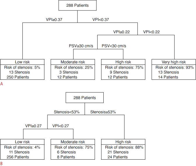
Results: In total, 288 patients (150 girls; median age at LT, 1.8 years [interquartile range, 0.9 to 3.6 years]) were included. Among the Doppler US parameters, venous pulsatility index (VPI) showed excellent diagnostic performance (area under the ROC curve [AUROC], 0.90; 95% confidence interval [CI], 0.86 to 0.93; Youden cut-off value, 0.40). Among the CT parameters, anastomotic site diameter (AUROC, 0.92; 95% CI, 0.88 to 0.95; Youden cut-off, 4.2 mm) and percentage of anastomotic site stenosis (AUROC, 0.88; 95% CI, 0.84 to 0.92; Youden cut-off, 35%) showed excellent and good diagnostic performance, respectively. A decision tree model combining the VPI, peak systolic velocity, and percentage of anastomotic site stenosis stratified patients according to the risk of hepatic outflow obstruction.
Conclusion: VPI, anastomotic site diameter, and percentage of anastomotic site stenosis were reliable imaging parameters for diagnosing hepatic outflow obstruction after pediatric LT using LL or LLS grafts.
UltrasonographyMedicine-Radiology, Nuclear Medicine and Imaging
CiteScore
5.10
自引率
6.50%
发文量
78
审稿时长
15 weeks
期刊介绍:
Ultrasonography, the official English-language journal of the Korean Society of Ultrasound in Medicine (KSUM), is an international peer-reviewed academic journal dedicated to practice, research, technology, and education dealing with medical ultrasound. It is renamed from the Journal of Korean Society of Ultrasound in Medicine in January 2014, and published four times per year: January 1, April 1, July 1, and October 1. Original articles, technical notes, topical reviews, perspectives, pictorial essays, and timely editorial materials are published in Ultrasonography covering state-of-the-art content.
Ultrasonography aims to provide updated information on new diagnostic concepts and technical developments, including experimental animal studies using new equipment in addition to well-designed reviews of contemporary issues in patient care. Along with running KSUM Open, the annual international congress of KSUM, Ultrasonography also serves as a medium for cooperation among physicians and specialists from around the world who are focusing on various ultrasound technology and disease problems and relevant basic science.
 求助内容:
求助内容: 应助结果提醒方式:


