{"title":"一例低级别子宫内膜基质肉瘤在核磁共振成像上表现为模仿子宫肌瘤的子宫内肿块。","authors":"Soichiro Tamada, Hiromi Edo, Taishi Sakima, Ryo Tanaka, Kohei Shikata, Soko Nishitani, Morikazu Miyamoto, Masashi Takano, Keisuke Kuboshima, Kosuke Miyai, Sho Ogata, Hiroshi Shinmoto","doi":"10.1093/bjrcr/uaad012","DOIUrl":null,"url":null,"abstract":"<p><p>A low-grade endometrial stromal sarcoma (ESS) has a pattern of presenting as an intramyometrial mass and is often misdiagnosed as cellular leiomyoma or degenerative uterine leiomyoma. A low-grade ESS is a malignant tumour that requires total hysterectomy with bilateral salpingo-oophorectomy; while a leiomyoma is a benign tumour and could be acceptable for enucleation. As the treatment strategies differ between a low-grade ESS and leiomyoma, radiologists should be familiar with the characteristic MRI findings of a low-grade ESS. A 51-year-old woman with abnormal uterine bleeding had been observed for 2 years at a previous hospital for a uterine leiomyoma based on MRI findings. A contrast-enhanced MRI demonstrated an intramyometrial mass composed of three components with the hypointense rim on T2-weighted images (T2WI): the first component was a homogeneous solid structure with mild hyperintensity on T2WI with a low apparent diffusion coefficient value; the second component was cystic; the third component was a structure of low signal intensity on T2WI similar to the muscle. Although a degenerative uterine leiomyoma was a differential diagnosis, these MRI findings were suggestive of a low-grade ESS. A total abdominal hysterectomy, bilateral salpingo-oophorectomy, pelvic lymphadenectomy, and partial omentectomy were performed. The pathological diagnosis was a low-grade ESS. In a low-grade ESS, there are three major patterns of MRI findings: one of these patterns is the less popular but clinically important intramyometrial mass pattern, which can be misdiagnosed as a leiomyoma, and this case conformed to this pattern.</p>","PeriodicalId":45216,"journal":{"name":"BJR Case Reports","volume":"10 1","pages":"uaad012"},"PeriodicalIF":0.5000,"publicationDate":"2023-12-18","publicationTypes":"Journal Article","fieldsOfStudy":null,"isOpenAccess":false,"openAccessPdf":"https://www.ncbi.nlm.nih.gov/pmc/articles/PMC10860526/pdf/","citationCount":"0","resultStr":"{\"title\":\"A case of low-grade endometrial stromal sarcoma presented as an intramyometrial mass mimicking uterine leiomyoma on MRI.\",\"authors\":\"Soichiro Tamada, Hiromi Edo, Taishi Sakima, Ryo Tanaka, Kohei Shikata, Soko Nishitani, Morikazu Miyamoto, Masashi Takano, Keisuke Kuboshima, Kosuke Miyai, Sho Ogata, Hiroshi Shinmoto\",\"doi\":\"10.1093/bjrcr/uaad012\",\"DOIUrl\":null,\"url\":null,\"abstract\":\"<p><p>A low-grade endometrial stromal sarcoma (ESS) has a pattern of presenting as an intramyometrial mass and is often misdiagnosed as cellular leiomyoma or degenerative uterine leiomyoma. A low-grade ESS is a malignant tumour that requires total hysterectomy with bilateral salpingo-oophorectomy; while a leiomyoma is a benign tumour and could be acceptable for enucleation. As the treatment strategies differ between a low-grade ESS and leiomyoma, radiologists should be familiar with the characteristic MRI findings of a low-grade ESS. A 51-year-old woman with abnormal uterine bleeding had been observed for 2 years at a previous hospital for a uterine leiomyoma based on MRI findings. A contrast-enhanced MRI demonstrated an intramyometrial mass composed of three components with the hypointense rim on T2-weighted images (T2WI): the first component was a homogeneous solid structure with mild hyperintensity on T2WI with a low apparent diffusion coefficient value; the second component was cystic; the third component was a structure of low signal intensity on T2WI similar to the muscle. Although a degenerative uterine leiomyoma was a differential diagnosis, these MRI findings were suggestive of a low-grade ESS. A total abdominal hysterectomy, bilateral salpingo-oophorectomy, pelvic lymphadenectomy, and partial omentectomy were performed. The pathological diagnosis was a low-grade ESS. In a low-grade ESS, there are three major patterns of MRI findings: one of these patterns is the less popular but clinically important intramyometrial mass pattern, which can be misdiagnosed as a leiomyoma, and this case conformed to this pattern.</p>\",\"PeriodicalId\":45216,\"journal\":{\"name\":\"BJR Case Reports\",\"volume\":\"10 1\",\"pages\":\"uaad012\"},\"PeriodicalIF\":0.5000,\"publicationDate\":\"2023-12-18\",\"publicationTypes\":\"Journal Article\",\"fieldsOfStudy\":null,\"isOpenAccess\":false,\"openAccessPdf\":\"https://www.ncbi.nlm.nih.gov/pmc/articles/PMC10860526/pdf/\",\"citationCount\":\"0\",\"resultStr\":null,\"platform\":\"Semanticscholar\",\"paperid\":null,\"PeriodicalName\":\"BJR Case Reports\",\"FirstCategoryId\":\"1085\",\"ListUrlMain\":\"https://doi.org/10.1093/bjrcr/uaad012\",\"RegionNum\":0,\"RegionCategory\":null,\"ArticlePicture\":[],\"TitleCN\":null,\"AbstractTextCN\":null,\"PMCID\":null,\"EPubDate\":\"2024/1/1 0:00:00\",\"PubModel\":\"eCollection\",\"JCR\":\"Q4\",\"JCRName\":\"RADIOLOGY, NUCLEAR MEDICINE & MEDICAL IMAGING\",\"Score\":null,\"Total\":0}","platform":"Semanticscholar","paperid":null,"PeriodicalName":"BJR Case Reports","FirstCategoryId":"1085","ListUrlMain":"https://doi.org/10.1093/bjrcr/uaad012","RegionNum":0,"RegionCategory":null,"ArticlePicture":[],"TitleCN":null,"AbstractTextCN":null,"PMCID":null,"EPubDate":"2024/1/1 0:00:00","PubModel":"eCollection","JCR":"Q4","JCRName":"RADIOLOGY, NUCLEAR MEDICINE & MEDICAL IMAGING","Score":null,"Total":0}
A case of low-grade endometrial stromal sarcoma presented as an intramyometrial mass mimicking uterine leiomyoma on MRI.
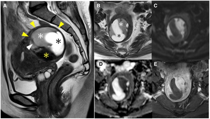
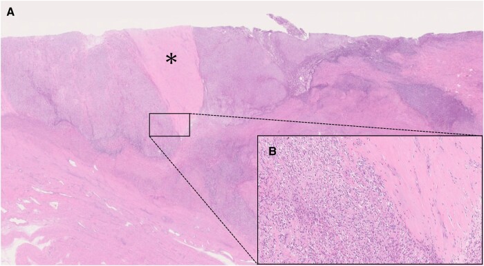
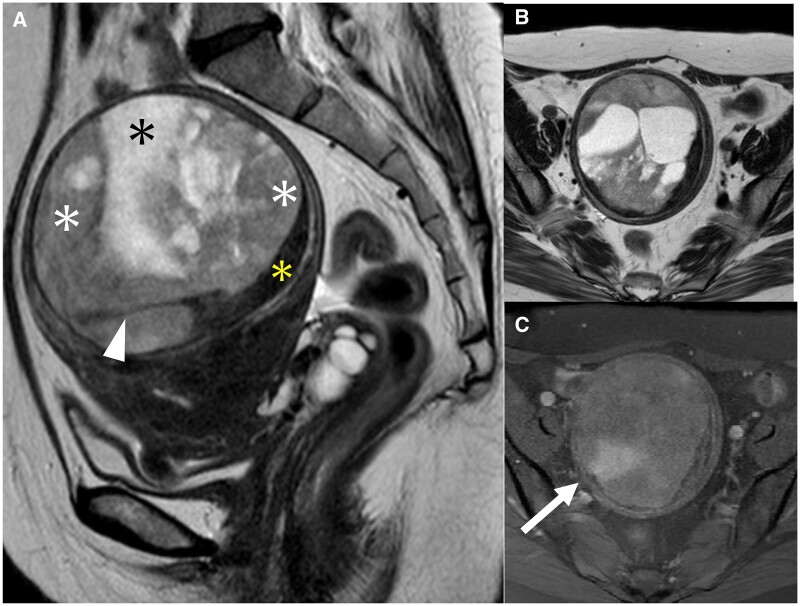
A low-grade endometrial stromal sarcoma (ESS) has a pattern of presenting as an intramyometrial mass and is often misdiagnosed as cellular leiomyoma or degenerative uterine leiomyoma. A low-grade ESS is a malignant tumour that requires total hysterectomy with bilateral salpingo-oophorectomy; while a leiomyoma is a benign tumour and could be acceptable for enucleation. As the treatment strategies differ between a low-grade ESS and leiomyoma, radiologists should be familiar with the characteristic MRI findings of a low-grade ESS. A 51-year-old woman with abnormal uterine bleeding had been observed for 2 years at a previous hospital for a uterine leiomyoma based on MRI findings. A contrast-enhanced MRI demonstrated an intramyometrial mass composed of three components with the hypointense rim on T2-weighted images (T2WI): the first component was a homogeneous solid structure with mild hyperintensity on T2WI with a low apparent diffusion coefficient value; the second component was cystic; the third component was a structure of low signal intensity on T2WI similar to the muscle. Although a degenerative uterine leiomyoma was a differential diagnosis, these MRI findings were suggestive of a low-grade ESS. A total abdominal hysterectomy, bilateral salpingo-oophorectomy, pelvic lymphadenectomy, and partial omentectomy were performed. The pathological diagnosis was a low-grade ESS. In a low-grade ESS, there are three major patterns of MRI findings: one of these patterns is the less popular but clinically important intramyometrial mass pattern, which can be misdiagnosed as a leiomyoma, and this case conformed to this pattern.

 求助内容:
求助内容: 应助结果提醒方式:


