{"title":"替换大脑后动脉","authors":"Hideki Endo, Kohei Ishikawa, Hidetoshi Ono, Kaori Honjo, Hirohiko Nakamura","doi":"10.1007/s00276-023-03294-6","DOIUrl":null,"url":null,"abstract":"<h3 data-test=\"abstract-sub-heading\">Purpose</h3><p>Replaced posterior cerebral artery (PCA), defined as a hyperplastic anterior choroidal artery (AChA) supplying all branches of the PCA, is an extremely rare anatomical variation. To the best of our knowledge, there are only a few reports of replaced PCA.</p><h3 data-test=\"abstract-sub-heading\">Methods</h3><p>Herein, we report a case of replaced PCA diagnosed by digital subtraction angiography.</p><h3 data-test=\"abstract-sub-heading\">Results</h3><p>A 76-year-old woman visited a neurosurgical clinic because of headache and vertigo. Magnetic resonance imaging and magnetic resonance angiography incidentally revealed a left internal carotid artery aneurysm. She was referred to our hospital for further examination and treatment of the unruptured intracranial aneurysm. Left internal carotid angiography revealed a paraclinoid aneurysm. We also incidentally found an anomalous hyperplastic AChA distal to the aneurysm. This hyperplastic AChA supplied not only the AChA territory but also the entire PCA territory. No vessels that could be a normal AChA or posterior communicating artery were identified along the left internal carotid artery. Vertebral angiography demonstrated that the left PCA was not visualized. With these findings, we diagnosed anomalous hyperplastic AChoA in this case as replaced PCA.</p><h3 data-test=\"abstract-sub-heading\">Conclusion</h3><p>Careful imaging assessment is important to identify replaced PCA. Both direct findings of a hyperplastic AChA course and perfusion territory and indirect findings of the absence of the original PCA are useful in the diagnosis of replaced PCA.</p>","PeriodicalId":49296,"journal":{"name":"Surgical and Radiologic Anatomy","volume":"37 1","pages":""},"PeriodicalIF":1.2000,"publicationDate":"2024-02-05","publicationTypes":"Journal Article","fieldsOfStudy":null,"isOpenAccess":false,"openAccessPdf":"","citationCount":"0","resultStr":"{\"title\":\"Replaced posterior cerebral artery\",\"authors\":\"Hideki Endo, Kohei Ishikawa, Hidetoshi Ono, Kaori Honjo, Hirohiko Nakamura\",\"doi\":\"10.1007/s00276-023-03294-6\",\"DOIUrl\":null,\"url\":null,\"abstract\":\"<h3 data-test=\\\"abstract-sub-heading\\\">Purpose</h3><p>Replaced posterior cerebral artery (PCA), defined as a hyperplastic anterior choroidal artery (AChA) supplying all branches of the PCA, is an extremely rare anatomical variation. To the best of our knowledge, there are only a few reports of replaced PCA.</p><h3 data-test=\\\"abstract-sub-heading\\\">Methods</h3><p>Herein, we report a case of replaced PCA diagnosed by digital subtraction angiography.</p><h3 data-test=\\\"abstract-sub-heading\\\">Results</h3><p>A 76-year-old woman visited a neurosurgical clinic because of headache and vertigo. Magnetic resonance imaging and magnetic resonance angiography incidentally revealed a left internal carotid artery aneurysm. She was referred to our hospital for further examination and treatment of the unruptured intracranial aneurysm. Left internal carotid angiography revealed a paraclinoid aneurysm. We also incidentally found an anomalous hyperplastic AChA distal to the aneurysm. This hyperplastic AChA supplied not only the AChA territory but also the entire PCA territory. No vessels that could be a normal AChA or posterior communicating artery were identified along the left internal carotid artery. Vertebral angiography demonstrated that the left PCA was not visualized. With these findings, we diagnosed anomalous hyperplastic AChoA in this case as replaced PCA.</p><h3 data-test=\\\"abstract-sub-heading\\\">Conclusion</h3><p>Careful imaging assessment is important to identify replaced PCA. Both direct findings of a hyperplastic AChA course and perfusion territory and indirect findings of the absence of the original PCA are useful in the diagnosis of replaced PCA.</p>\",\"PeriodicalId\":49296,\"journal\":{\"name\":\"Surgical and Radiologic Anatomy\",\"volume\":\"37 1\",\"pages\":\"\"},\"PeriodicalIF\":1.2000,\"publicationDate\":\"2024-02-05\",\"publicationTypes\":\"Journal Article\",\"fieldsOfStudy\":null,\"isOpenAccess\":false,\"openAccessPdf\":\"\",\"citationCount\":\"0\",\"resultStr\":null,\"platform\":\"Semanticscholar\",\"paperid\":null,\"PeriodicalName\":\"Surgical and Radiologic Anatomy\",\"FirstCategoryId\":\"3\",\"ListUrlMain\":\"https://doi.org/10.1007/s00276-023-03294-6\",\"RegionNum\":4,\"RegionCategory\":\"医学\",\"ArticlePicture\":[],\"TitleCN\":null,\"AbstractTextCN\":null,\"PMCID\":null,\"EPubDate\":\"\",\"PubModel\":\"\",\"JCR\":\"Q3\",\"JCRName\":\"ANATOMY & MORPHOLOGY\",\"Score\":null,\"Total\":0}","platform":"Semanticscholar","paperid":null,"PeriodicalName":"Surgical and Radiologic Anatomy","FirstCategoryId":"3","ListUrlMain":"https://doi.org/10.1007/s00276-023-03294-6","RegionNum":4,"RegionCategory":"医学","ArticlePicture":[],"TitleCN":null,"AbstractTextCN":null,"PMCID":null,"EPubDate":"","PubModel":"","JCR":"Q3","JCRName":"ANATOMY & MORPHOLOGY","Score":null,"Total":0}
Replaced posterior cerebral artery (PCA), defined as a hyperplastic anterior choroidal artery (AChA) supplying all branches of the PCA, is an extremely rare anatomical variation. To the best of our knowledge, there are only a few reports of replaced PCA.
Methods
Herein, we report a case of replaced PCA diagnosed by digital subtraction angiography.
Results
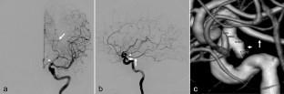
A 76-year-old woman visited a neurosurgical clinic because of headache and vertigo. Magnetic resonance imaging and magnetic resonance angiography incidentally revealed a left internal carotid artery aneurysm. She was referred to our hospital for further examination and treatment of the unruptured intracranial aneurysm. Left internal carotid angiography revealed a paraclinoid aneurysm. We also incidentally found an anomalous hyperplastic AChA distal to the aneurysm. This hyperplastic AChA supplied not only the AChA territory but also the entire PCA territory. No vessels that could be a normal AChA or posterior communicating artery were identified along the left internal carotid artery. Vertebral angiography demonstrated that the left PCA was not visualized. With these findings, we diagnosed anomalous hyperplastic AChoA in this case as replaced PCA.
Conclusion
Careful imaging assessment is important to identify replaced PCA. Both direct findings of a hyperplastic AChA course and perfusion territory and indirect findings of the absence of the original PCA are useful in the diagnosis of replaced PCA.
期刊介绍:
Anatomy is a morphological science which cannot fail to interest the clinician. The practical application of anatomical research to clinical problems necessitates special adaptation and selectivity in choosing from numerous international works. Although there is a tendency to believe that meaningful advances in anatomy are unlikely, constant revision is necessary. Surgical and Radiologic Anatomy, the first international journal of Clinical anatomy has been created in this spirit.
Its goal is to serve clinicians, regardless of speciality-physicians, surgeons, radiologists or other specialists-as an indispensable aid with which they can improve their knowledge of anatomy. Each issue includes: Original papers, review articles, articles on the anatomical bases of medical, surgical and radiological techniques, articles of normal radiologic anatomy, brief reviews of anatomical publications of clinical interest.
Particular attention is given to high quality illustrations, which are indispensable for a better understanding of anatomical problems.
Surgical and Radiologic Anatomy is a journal written by anatomists for clinicians with a special interest in anatomy.

 求助内容:
求助内容: 应助结果提醒方式:


