干细胞通过共培养减轻 PC12 细胞中由 OGD/R 介导的应激反应:通过 BDNF-TrkB 信号调节凋亡级联。

IF 3.3 3区 生物学 Q3 CELL BIOLOGY
Harpreet Kaur, Deepaneeta Sarmah, Aishika Datta, Anupom Borah, Dileep R Yavagal, Pallab Bhattacharya
{"title":"干细胞通过共培养减轻 PC12 细胞中由 OGD/R 介导的应激反应:通过 BDNF-TrkB 信号调节凋亡级联。","authors":"Harpreet Kaur, Deepaneeta Sarmah, Aishika Datta, Anupom Borah, Dileep R Yavagal, Pallab Bhattacharya","doi":"10.1007/s12192-022-01319-4","DOIUrl":null,"url":null,"abstract":"<p><p>Apoptosis mediated by endoplasmic reticulum (ER) stress plays a crucial role in several neurovascular disorders, including ischemia/reperfusion injury (I/R injury). Previous in vitro and in vivo studies have suggested that following I/R injury, ER stress is vital for mediating CCAT-enhancer-binding protein homologous protein (CHOP) and caspase-12-dependent apoptosis. However, its modulation in the presence of stem cells and the underlying mechanism of cytoprotection remains elusive. In vivo studies from our lab have reported that post-stroke endovascular administration of stem cells renders neuroprotection and regulates apoptosis mediated by ER stress. In the current study, a more robust in vitro validation has been undertaken to decipher the mechanism of stem cell-mediated cytoprotection. Results from our study have shown that oxygen-glucose deprivation/reoxygenation (OGD/R) potentiated ER stress and apoptosis in the pheochromocytoma 12 (PC12) cell line as evident by the increase of protein kinase R (PKR)-like ER kinase (p-PERK), p-Eukaryotic initiation factor 2α subunit (EIF2α), activation transcription factor 4 (ATF4), CHOP, and caspase 12 expressions. Following the co-culture of PC12 cells with MSCs, ER stress was significantly reduced, possibly via modulating the brain-derived neurotrophic factor (BDNF) signaling. Furthermore, inhibition of BDNF by inhibitor K252a abolished the protective effects of BDNF secreted by MSCs following OGD/R. Our study suggests that inhibition of ER stress-associated apoptotic pathway with MSCs co-culture following OGD/R may help to alleviate cellular injury and further substantiate the use of stem cells as a therapeutic modality toward neuroprotection following hypoxic injury or stroke in clinical settings.</p>","PeriodicalId":9684,"journal":{"name":"Cell Stress & Chaperones","volume":"28 6","pages":"1041-1051"},"PeriodicalIF":3.3000,"publicationDate":"2023-11-01","publicationTypes":"Journal Article","fieldsOfStudy":null,"isOpenAccess":false,"openAccessPdf":"","citationCount":"0","resultStr":"{\"title\":\"Stem cells alleviate OGD/R mediated stress response in PC12 cells following a co-culture: modulation of the apoptotic cascade through BDNF-TrkB signaling.\",\"authors\":\"Harpreet Kaur, Deepaneeta Sarmah, Aishika Datta, Anupom Borah, Dileep R Yavagal, Pallab Bhattacharya\",\"doi\":\"10.1007/s12192-022-01319-4\",\"DOIUrl\":null,\"url\":null,\"abstract\":\"<p><p>Apoptosis mediated by endoplasmic reticulum (ER) stress plays a crucial role in several neurovascular disorders, including ischemia/reperfusion injury (I/R injury). Previous in vitro and in vivo studies have suggested that following I/R injury, ER stress is vital for mediating CCAT-enhancer-binding protein homologous protein (CHOP) and caspase-12-dependent apoptosis. However, its modulation in the presence of stem cells and the underlying mechanism of cytoprotection remains elusive. In vivo studies from our lab have reported that post-stroke endovascular administration of stem cells renders neuroprotection and regulates apoptosis mediated by ER stress. In the current study, a more robust in vitro validation has been undertaken to decipher the mechanism of stem cell-mediated cytoprotection. Results from our study have shown that oxygen-glucose deprivation/reoxygenation (OGD/R) potentiated ER stress and apoptosis in the pheochromocytoma 12 (PC12) cell line as evident by the increase of protein kinase R (PKR)-like ER kinase (p-PERK), p-Eukaryotic initiation factor 2α subunit (EIF2α), activation transcription factor 4 (ATF4), CHOP, and caspase 12 expressions. Following the co-culture of PC12 cells with MSCs, ER stress was significantly reduced, possibly via modulating the brain-derived neurotrophic factor (BDNF) signaling. Furthermore, inhibition of BDNF by inhibitor K252a abolished the protective effects of BDNF secreted by MSCs following OGD/R. Our study suggests that inhibition of ER stress-associated apoptotic pathway with MSCs co-culture following OGD/R may help to alleviate cellular injury and further substantiate the use of stem cells as a therapeutic modality toward neuroprotection following hypoxic injury or stroke in clinical settings.</p>\",\"PeriodicalId\":9684,\"journal\":{\"name\":\"Cell Stress & Chaperones\",\"volume\":\"28 6\",\"pages\":\"1041-1051\"},\"PeriodicalIF\":3.3000,\"publicationDate\":\"2023-11-01\",\"publicationTypes\":\"Journal Article\",\"fieldsOfStudy\":null,\"isOpenAccess\":false,\"openAccessPdf\":\"\",\"citationCount\":\"0\",\"resultStr\":null,\"platform\":\"Semanticscholar\",\"paperid\":null,\"PeriodicalName\":\"Cell Stress & Chaperones\",\"FirstCategoryId\":\"99\",\"ListUrlMain\":\"https://doi.org/10.1007/s12192-022-01319-4\",\"RegionNum\":3,\"RegionCategory\":\"生物学\",\"ArticlePicture\":[],\"TitleCN\":null,\"AbstractTextCN\":null,\"PMCID\":null,\"EPubDate\":\"\",\"PubModel\":\"\",\"JCR\":\"Q3\",\"JCRName\":\"CELL BIOLOGY\",\"Score\":null,\"Total\":0}","platform":"Semanticscholar","paperid":null,"PeriodicalName":"Cell Stress & Chaperones","FirstCategoryId":"99","ListUrlMain":"https://doi.org/10.1007/s12192-022-01319-4","RegionNum":3,"RegionCategory":"生物学","ArticlePicture":[],"TitleCN":null,"AbstractTextCN":null,"PMCID":null,"EPubDate":"","PubModel":"","JCR":"Q3","JCRName":"CELL BIOLOGY","Score":null,"Total":0}
引用次数: 0

摘要

内质网(ER)应激介导的细胞凋亡在包括缺血/再灌注损伤(I/R 损伤)在内的多种神经血管疾病中起着至关重要的作用。之前的体外和体内研究表明,I/R 损伤后,ER 应激对于介导 CCAT-增强子结合蛋白同源蛋白(CHOP)和 caspase-12 依赖性凋亡至关重要。然而,干细胞存在时对ER应激的调控以及细胞保护的基本机制仍未确定。我们实验室的体内研究报告称,中风后血管内注射干细胞可提供神经保护,并调节由ER应激介导的细胞凋亡。在目前的研究中,我们进行了更有力的体外验证,以破译干细胞介导的细胞保护机制。我们的研究结果表明,氧-葡萄糖剥夺/复氧(OGD/R)增强了嗜铬细胞瘤12(PC12)细胞系的ER应激和细胞凋亡,表现为蛋白激酶R(PKR)-类ER激酶(p-PERK)、p-核糖体启动因子2α亚基(EIF2α)、活化转录因子4(ATF4)、CHOP和caspase 12表达的增加。PC12细胞与间充质干细胞共培养后,ER应激明显降低,这可能是通过调节脑源性神经营养因子(BDNF)信号传导实现的。此外,用抑制剂K252a抑制BDNF可消除间充质干细胞分泌的BDNF对OGD/R的保护作用。我们的研究表明,在OGD/R后与间充质干细胞共培养,抑制ER应激相关的细胞凋亡通路可能有助于减轻细胞损伤,并进一步证实了干细胞在缺氧损伤或中风后的临床神经保护治疗中的应用。
本文章由计算机程序翻译,如有差异,请以英文原文为准。
Stem cells alleviate OGD/R mediated stress response in PC12 cells following a co-culture: modulation of the apoptotic cascade through BDNF-TrkB signaling.

Apoptosis mediated by endoplasmic reticulum (ER) stress plays a crucial role in several neurovascular disorders, including ischemia/reperfusion injury (I/R injury). Previous in vitro and in vivo studies have suggested that following I/R injury, ER stress is vital for mediating CCAT-enhancer-binding protein homologous protein (CHOP) and caspase-12-dependent apoptosis. However, its modulation in the presence of stem cells and the underlying mechanism of cytoprotection remains elusive. In vivo studies from our lab have reported that post-stroke endovascular administration of stem cells renders neuroprotection and regulates apoptosis mediated by ER stress. In the current study, a more robust in vitro validation has been undertaken to decipher the mechanism of stem cell-mediated cytoprotection. Results from our study have shown that oxygen-glucose deprivation/reoxygenation (OGD/R) potentiated ER stress and apoptosis in the pheochromocytoma 12 (PC12) cell line as evident by the increase of protein kinase R (PKR)-like ER kinase (p-PERK), p-Eukaryotic initiation factor 2α subunit (EIF2α), activation transcription factor 4 (ATF4), CHOP, and caspase 12 expressions. Following the co-culture of PC12 cells with MSCs, ER stress was significantly reduced, possibly via modulating the brain-derived neurotrophic factor (BDNF) signaling. Furthermore, inhibition of BDNF by inhibitor K252a abolished the protective effects of BDNF secreted by MSCs following OGD/R. Our study suggests that inhibition of ER stress-associated apoptotic pathway with MSCs co-culture following OGD/R may help to alleviate cellular injury and further substantiate the use of stem cells as a therapeutic modality toward neuroprotection following hypoxic injury or stroke in clinical settings.

求助全文
通过发布文献求助,成功后即可免费获取论文全文。 去求助
来源期刊
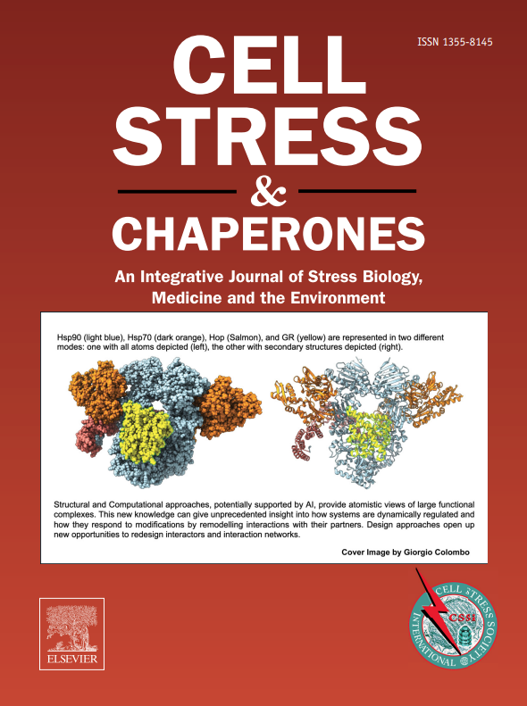
Cell Stress & Chaperones
Cell Stress & Chaperones 生物-细胞生物学
CiteScore
7.60
自引率
2.60%
发文量
59
审稿时长
6-12 weeks
期刊介绍: Cell Stress and Chaperones is an integrative journal that bridges the gap between laboratory model systems and natural populations. The journal captures the eclectic spirit of the cellular stress response field in a single, concentrated source of current information. Major emphasis is placed on the effects of climate change on individual species in the natural environment and their capacity to adapt. This emphasis expands our focus on stress biology and medicine by linking climate change effects to research on cellular stress responses of animals, micro-organisms and plants.
×
引用
GB/T 7714-2015
复制
MLA
复制
APA
复制
导出至
BibTeX EndNote RefMan NoteFirst NoteExpress
×
提示
您的信息不完整,为了账户安全,请先补充。
现在去补充
×
提示
您因"违规操作"
具体请查看互助需知
我知道了
×
提示
确定
请完成安全验证×
copy
已复制链接
快去分享给好友吧!
我知道了
右上角分享
点击右上角分享
0
联系我们:info@booksci.cn Book学术提供免费学术资源搜索服务,方便国内外学者检索中英文文献。致力于提供最便捷和优质的服务体验。 Copyright © 2023 布克学术 All rights reserved.
京ICP备2023020795号-1
ghs 京公网安备 11010802042870号
Book学术文献互助
Book学术文献互助群
群 号:481959085
Book学术官方微信