{"title":"溶酶体靶向嵌合体(LYTAC):靶向降解致癌膜蛋白的银弹","authors":"Qingquan Zheng, Jiawei Guo, Rui Ma, Wenchen Pu","doi":"10.1002/mog2.64","DOIUrl":null,"url":null,"abstract":"<p>Recently, the group of Prof. Carolyn Bertozzi, a laureate of the Nobel Prize in chemistry 2022, reported the detailed mechanism of lysosome-targeting chimera (LYTAC) in the journal of <i>Science</i>,<span><sup>1</sup></span> after the publication of their first LYTAC molecule in <i>Nature</i> in 2020.<span><sup>2</sup></span> The establishment of LYTAC, a subtype of targeted protein degradation technology, expands the scope of protein degradation to extracellular and membrane-associated targets, and Bertozzi group's new discovery is expected to accelerate the development of LYTAC in cancer therapy.</p><p>Cell membranes play a critical role in various cellular processes, including signaling transduction, cell adhesion, transport of biomolecules and immunity. Proteins embedded in or associated with the cell membrane are key executants of the function of cell membrane, and their dysregulation contributes to tumorigenesis and development of human cancers.<span><sup>3</sup></span> For example, epidermal growth factor receptor (EGFR) is a receptor tyrosine kinase for epithelial growth factor (EGF) and transforming growth factor α (TGF-α), belonging to the ErbB receptor family. Activation of EGFR signaling promotes cell proliferation, survival, angiogenesis and metastasis of diverse malignancies.<span><sup>3</sup></span> Moreover, hepatocyte growth factor receptor (c-Met, HGFR) is another oncogenic receptor tyrosine kinase in diverse cancers. Upon the binding to hepatocyte growth factor (HGF), c-Met is activated via autophosphorylation, leading to the initiation of oncogenic downstream signaling cascades, such as PI3K/AKT and RAS/ERK pathways.<span><sup>3</sup></span> Given their central role in cancer-promoting processes, EGFR and c-Met has become attractive targets for cancer therapies. Small-molecule tyrosine kinase inhibitors (EGFR: gefitinib, afatinib, osimertinib, etc.; c-Met: capmatinib, tepotinib, savolitinib, etc.) and monoclonal antibodies (EGFR: cetuximab, panitumumab; EGFR/c-Met: amivantamab; c-Met: emibetuzumab), have been developed and approved for the treatment of various cancers, including lung and colorectal cancers (Figure 1A). But severe acquired resistance (e.g., via EGFR mutations) and limited therapeutic efficacy (slightly prolonged overall survival) of these treatments restrict their clinical benefit for patients. Moreover, nonenzymatic function of membrane proteins, such as protein–protein interactions, could not be interfered with by kinase inhibitors or monoclonal antibodies, calling for new strategies to control these oncogenic membrane proteins.</p><p>Targeted protein degradation (TPD) is a therapeutic approach that aims to selectively remove disease-causing or undesirable proteins from cells by inducing their degradation, with multiple therapies entering clinical trials and targeting proteins that are previously considered “undruggable.”<span><sup>4</sup></span> There are two main protein degradation mechanisms within cells, including ubiquitin-proteasome system (UPS) and lysosome pathway. By triggering chemically induced proximity to form ternary complex assembly via heterobifunctional molecules, various TPD techniques have been developed based on proteasome (PROTAC, molecular glue, etc.), lysosome (LYTAC, AUTAC, ATTEC, etc.), or both (PROTAB).<span><sup>4</sup></span> Currently, some membrane proteins could be degraded through TPD technologies. For example, Jang et al. developed an allosteric EGFR proteolysis-targeting chimera (PROTAC), DDC-01-163, which with selective activity against various clinically relevant EGFR mutants (L858R/T790M) as a single agent or combined with an ATP-site inhibitor osimertinib (Figure 1B, left).<span><sup>5</sup></span> Recently, Marei et al. reported proteolysis-targeting antibodies (PROTABs, a class of bispecific antibodies) that tethered cell-surface E3 ubiquitin ligases (RNF43 or ZNRF3) to transmembrane proteins (IGF1R, HER2, and PD-L1) for targeted degradation via both UPS and lysosome pathway, providing a strategy for the rapid development of potent, bioavailable and tissue-selective degraders of membrane proteins.<span><sup>6</sup></span></p><p>In 2020, Bertozzi group prepared bifunctional molecules that consisted of antibodies conjugated to chemically synthesized glycopeptide ligands, the agonists of the cation-independent mannose-6-phosphate receptor (CI-M6PR, a cell-surface lysosome-shuttling receptor). These conjugates recognized both CI-M6PR and the extracellular domain of target proteins, inducing the lysosome-mediated targeted degradation of membrane proteins, such as EGFR, CD71, programmed death-ligand 1 (PD-L1) and apolipoprotein E4. Notably, the data from CRISPR interference screen suggested an involvement of CI-M6PR-mediated cargo internalization in cell lines, and uncovered the participation of exocyst complex in these processes. Thus, these evidence demonstrated the feasibility of degrading membrane proteins through lysosomes by activating chemically induced proximity. Thus, this technique was termed as lysosome-targeting chimaeras (LYTACs).<span><sup>2</sup></span> However, the cellular characteristics that regulate the behavior of LYTACs to hijack lysosome machinery for membrane protein degradation are largely unknown. There is an urgent need for identifying the cellular determinants that modulate the efficacy of LYTACs-induced lysosomal degradation, facilitating the understanding of their molecular and cellular mechanisms.</p><p>To this end, Bertozzi group subsequently performed an unbiased genome-wide CRISPR knockout screening approach complemented by proteomics to map the key regulators of LYTAC-mediated membrane protein degradation in human cells. The results indicated that inhibiting retromer genes (e.g., <i>VPS35</i>, <i>SNX3</i>, <i>VPS29</i>, and <i>VPS26A</i>) to reduce LYTAC recycling enhanced the target degradation. Moreover, genes that involved in cullin3 (CUL3) neddylation, such as <i>CUL3</i>, <i>UBA3</i>, and <i>CAND1</i>, promoted the E3 ligase activity as well as the transport of LYTAC-target protein complexes to lysosomes. Thereby, levels of neddylated CUL3 could act as a predictive marker for LYTAC efficacy. Additionally, membrane CI-M6PR receptors were partially engaged by endogenous mannose 6-phosphate (M6P)-modified lysosomal glycoproteins. Blockage of M6P biosynthesis genes (e.g., <i>ALGO12</i>, <i>GNPTAB</i>) upregulated the ratio of unoccupied receptors, increasing LYTAC-receptor internalization and the degradation of cell surface proteins, including EGFR and c-Met.<span><sup>1</sup></span> Overall, this work discovered a series of critical cellular regulators that modulated LYTAC-mediated degradation of EGFR and c-Met (Figure 1B, right), giving important support for understanding LYTAC mechanism and developing next-generation LYTAC with improved clinical potential.</p><p>With the development and maturity of monoclonal antibody technology based on phage display, hybridoma cell and single B cell techniques, discovery and optimization of specific monoclonal antibodies is highly efficient. Furthermore, the synthesis and characterization of LYTAC could follow the route of antibody-drug conjugates (ADCs), because they share similar chemical compositions. Therefore, LYTAC is highly promising as a general platform for targeting extracellular and membrane proteins, the products of 40% of all protein-encoding genes. However, for targeted therapy of cancers, the following issues still need to be considered: Comprehensive in vivo evidence is needed to support the pharmacodynamics, pharmacokinetics and safety of LYTAC as a treatment option; Cooperative diagnosis is necessary for characterizing target protein, predictive biomarkers and LYTAC-related regulators to confirm suitable cancer patients, and for determining whether combination therapy is needed, such as in combination with the inhibitors of <i>ALGO12</i> or <i>GNPTAB</i>; Mutations of many oncogenic membrane proteins lead to tumor heterogeneity and/or drug resistance, and it is still uncertain whether LYTAC could overcome or bypass the negative effect of protein mutations. Since 2020, companies, such as Lycia Therapeutics (founded by Prof. Bertozzi) and Avilar Therapeutics, have undergone translational research around LYTAC technology. We look forward to these new discoveries driving LYTAC into novel therapies that benefit patients.</p><p>All authors were involved in the writing of the manuscript. Qingquan Zheng and Wenchen Pu initiated the conception and outline. Qingquan Zheng, Jiawei Guo, and Wenchen Pu organized and processed the figure. Jiawei Guo and Rui Ma revised manuscript. Jiawei Guo and Wenchen Pu were involved in study supervision. All authors have read and approved the final manuscript.</p><p>The authors declare no conflict of interest.</p><p>The authors declare that human ethics approval was not needed for this highlight.</p>","PeriodicalId":100902,"journal":{"name":"MedComm – Oncology","volume":"3 1","pages":""},"PeriodicalIF":0.0000,"publicationDate":"2024-01-14","publicationTypes":"Journal Article","fieldsOfStudy":null,"isOpenAccess":false,"openAccessPdf":"https://onlinelibrary.wiley.com/doi/epdf/10.1002/mog2.64","citationCount":"0","resultStr":"{\"title\":\"Lysosome-targeting chimera (LYTAC): A silver bullet for targeted degradation of oncogenic membrane proteins\",\"authors\":\"Qingquan Zheng, Jiawei Guo, Rui Ma, Wenchen Pu\",\"doi\":\"10.1002/mog2.64\",\"DOIUrl\":null,\"url\":null,\"abstract\":\"<p>Recently, the group of Prof. Carolyn Bertozzi, a laureate of the Nobel Prize in chemistry 2022, reported the detailed mechanism of lysosome-targeting chimera (LYTAC) in the journal of <i>Science</i>,<span><sup>1</sup></span> after the publication of their first LYTAC molecule in <i>Nature</i> in 2020.<span><sup>2</sup></span> The establishment of LYTAC, a subtype of targeted protein degradation technology, expands the scope of protein degradation to extracellular and membrane-associated targets, and Bertozzi group's new discovery is expected to accelerate the development of LYTAC in cancer therapy.</p><p>Cell membranes play a critical role in various cellular processes, including signaling transduction, cell adhesion, transport of biomolecules and immunity. Proteins embedded in or associated with the cell membrane are key executants of the function of cell membrane, and their dysregulation contributes to tumorigenesis and development of human cancers.<span><sup>3</sup></span> For example, epidermal growth factor receptor (EGFR) is a receptor tyrosine kinase for epithelial growth factor (EGF) and transforming growth factor α (TGF-α), belonging to the ErbB receptor family. Activation of EGFR signaling promotes cell proliferation, survival, angiogenesis and metastasis of diverse malignancies.<span><sup>3</sup></span> Moreover, hepatocyte growth factor receptor (c-Met, HGFR) is another oncogenic receptor tyrosine kinase in diverse cancers. Upon the binding to hepatocyte growth factor (HGF), c-Met is activated via autophosphorylation, leading to the initiation of oncogenic downstream signaling cascades, such as PI3K/AKT and RAS/ERK pathways.<span><sup>3</sup></span> Given their central role in cancer-promoting processes, EGFR and c-Met has become attractive targets for cancer therapies. Small-molecule tyrosine kinase inhibitors (EGFR: gefitinib, afatinib, osimertinib, etc.; c-Met: capmatinib, tepotinib, savolitinib, etc.) and monoclonal antibodies (EGFR: cetuximab, panitumumab; EGFR/c-Met: amivantamab; c-Met: emibetuzumab), have been developed and approved for the treatment of various cancers, including lung and colorectal cancers (Figure 1A). But severe acquired resistance (e.g., via EGFR mutations) and limited therapeutic efficacy (slightly prolonged overall survival) of these treatments restrict their clinical benefit for patients. Moreover, nonenzymatic function of membrane proteins, such as protein–protein interactions, could not be interfered with by kinase inhibitors or monoclonal antibodies, calling for new strategies to control these oncogenic membrane proteins.</p><p>Targeted protein degradation (TPD) is a therapeutic approach that aims to selectively remove disease-causing or undesirable proteins from cells by inducing their degradation, with multiple therapies entering clinical trials and targeting proteins that are previously considered “undruggable.”<span><sup>4</sup></span> There are two main protein degradation mechanisms within cells, including ubiquitin-proteasome system (UPS) and lysosome pathway. By triggering chemically induced proximity to form ternary complex assembly via heterobifunctional molecules, various TPD techniques have been developed based on proteasome (PROTAC, molecular glue, etc.), lysosome (LYTAC, AUTAC, ATTEC, etc.), or both (PROTAB).<span><sup>4</sup></span> Currently, some membrane proteins could be degraded through TPD technologies. For example, Jang et al. developed an allosteric EGFR proteolysis-targeting chimera (PROTAC), DDC-01-163, which with selective activity against various clinically relevant EGFR mutants (L858R/T790M) as a single agent or combined with an ATP-site inhibitor osimertinib (Figure 1B, left).<span><sup>5</sup></span> Recently, Marei et al. reported proteolysis-targeting antibodies (PROTABs, a class of bispecific antibodies) that tethered cell-surface E3 ubiquitin ligases (RNF43 or ZNRF3) to transmembrane proteins (IGF1R, HER2, and PD-L1) for targeted degradation via both UPS and lysosome pathway, providing a strategy for the rapid development of potent, bioavailable and tissue-selective degraders of membrane proteins.<span><sup>6</sup></span></p><p>In 2020, Bertozzi group prepared bifunctional molecules that consisted of antibodies conjugated to chemically synthesized glycopeptide ligands, the agonists of the cation-independent mannose-6-phosphate receptor (CI-M6PR, a cell-surface lysosome-shuttling receptor). These conjugates recognized both CI-M6PR and the extracellular domain of target proteins, inducing the lysosome-mediated targeted degradation of membrane proteins, such as EGFR, CD71, programmed death-ligand 1 (PD-L1) and apolipoprotein E4. Notably, the data from CRISPR interference screen suggested an involvement of CI-M6PR-mediated cargo internalization in cell lines, and uncovered the participation of exocyst complex in these processes. Thus, these evidence demonstrated the feasibility of degrading membrane proteins through lysosomes by activating chemically induced proximity. Thus, this technique was termed as lysosome-targeting chimaeras (LYTACs).<span><sup>2</sup></span> However, the cellular characteristics that regulate the behavior of LYTACs to hijack lysosome machinery for membrane protein degradation are largely unknown. There is an urgent need for identifying the cellular determinants that modulate the efficacy of LYTACs-induced lysosomal degradation, facilitating the understanding of their molecular and cellular mechanisms.</p><p>To this end, Bertozzi group subsequently performed an unbiased genome-wide CRISPR knockout screening approach complemented by proteomics to map the key regulators of LYTAC-mediated membrane protein degradation in human cells. The results indicated that inhibiting retromer genes (e.g., <i>VPS35</i>, <i>SNX3</i>, <i>VPS29</i>, and <i>VPS26A</i>) to reduce LYTAC recycling enhanced the target degradation. Moreover, genes that involved in cullin3 (CUL3) neddylation, such as <i>CUL3</i>, <i>UBA3</i>, and <i>CAND1</i>, promoted the E3 ligase activity as well as the transport of LYTAC-target protein complexes to lysosomes. Thereby, levels of neddylated CUL3 could act as a predictive marker for LYTAC efficacy. Additionally, membrane CI-M6PR receptors were partially engaged by endogenous mannose 6-phosphate (M6P)-modified lysosomal glycoproteins. Blockage of M6P biosynthesis genes (e.g., <i>ALGO12</i>, <i>GNPTAB</i>) upregulated the ratio of unoccupied receptors, increasing LYTAC-receptor internalization and the degradation of cell surface proteins, including EGFR and c-Met.<span><sup>1</sup></span> Overall, this work discovered a series of critical cellular regulators that modulated LYTAC-mediated degradation of EGFR and c-Met (Figure 1B, right), giving important support for understanding LYTAC mechanism and developing next-generation LYTAC with improved clinical potential.</p><p>With the development and maturity of monoclonal antibody technology based on phage display, hybridoma cell and single B cell techniques, discovery and optimization of specific monoclonal antibodies is highly efficient. Furthermore, the synthesis and characterization of LYTAC could follow the route of antibody-drug conjugates (ADCs), because they share similar chemical compositions. Therefore, LYTAC is highly promising as a general platform for targeting extracellular and membrane proteins, the products of 40% of all protein-encoding genes. However, for targeted therapy of cancers, the following issues still need to be considered: Comprehensive in vivo evidence is needed to support the pharmacodynamics, pharmacokinetics and safety of LYTAC as a treatment option; Cooperative diagnosis is necessary for characterizing target protein, predictive biomarkers and LYTAC-related regulators to confirm suitable cancer patients, and for determining whether combination therapy is needed, such as in combination with the inhibitors of <i>ALGO12</i> or <i>GNPTAB</i>; Mutations of many oncogenic membrane proteins lead to tumor heterogeneity and/or drug resistance, and it is still uncertain whether LYTAC could overcome or bypass the negative effect of protein mutations. Since 2020, companies, such as Lycia Therapeutics (founded by Prof. Bertozzi) and Avilar Therapeutics, have undergone translational research around LYTAC technology. We look forward to these new discoveries driving LYTAC into novel therapies that benefit patients.</p><p>All authors were involved in the writing of the manuscript. Qingquan Zheng and Wenchen Pu initiated the conception and outline. Qingquan Zheng, Jiawei Guo, and Wenchen Pu organized and processed the figure. Jiawei Guo and Rui Ma revised manuscript. Jiawei Guo and Wenchen Pu were involved in study supervision. All authors have read and approved the final manuscript.</p><p>The authors declare no conflict of interest.</p><p>The authors declare that human ethics approval was not needed for this highlight.</p>\",\"PeriodicalId\":100902,\"journal\":{\"name\":\"MedComm – Oncology\",\"volume\":\"3 1\",\"pages\":\"\"},\"PeriodicalIF\":0.0000,\"publicationDate\":\"2024-01-14\",\"publicationTypes\":\"Journal Article\",\"fieldsOfStudy\":null,\"isOpenAccess\":false,\"openAccessPdf\":\"https://onlinelibrary.wiley.com/doi/epdf/10.1002/mog2.64\",\"citationCount\":\"0\",\"resultStr\":null,\"platform\":\"Semanticscholar\",\"paperid\":null,\"PeriodicalName\":\"MedComm – Oncology\",\"FirstCategoryId\":\"1085\",\"ListUrlMain\":\"https://onlinelibrary.wiley.com/doi/10.1002/mog2.64\",\"RegionNum\":0,\"RegionCategory\":null,\"ArticlePicture\":[],\"TitleCN\":null,\"AbstractTextCN\":null,\"PMCID\":null,\"EPubDate\":\"\",\"PubModel\":\"\",\"JCR\":\"\",\"JCRName\":\"\",\"Score\":null,\"Total\":0}","platform":"Semanticscholar","paperid":null,"PeriodicalName":"MedComm – Oncology","FirstCategoryId":"1085","ListUrlMain":"https://onlinelibrary.wiley.com/doi/10.1002/mog2.64","RegionNum":0,"RegionCategory":null,"ArticlePicture":[],"TitleCN":null,"AbstractTextCN":null,"PMCID":null,"EPubDate":"","PubModel":"","JCR":"","JCRName":"","Score":null,"Total":0}
Lysosome-targeting chimera (LYTAC): A silver bullet for targeted degradation of oncogenic membrane proteins
Recently, the group of Prof. Carolyn Bertozzi, a laureate of the Nobel Prize in chemistry 2022, reported the detailed mechanism of lysosome-targeting chimera (LYTAC) in the journal of Science,1 after the publication of their first LYTAC molecule in Nature in 2020.2 The establishment of LYTAC, a subtype of targeted protein degradation technology, expands the scope of protein degradation to extracellular and membrane-associated targets, and Bertozzi group's new discovery is expected to accelerate the development of LYTAC in cancer therapy.
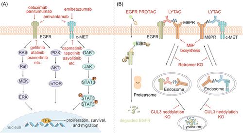
Cell membranes play a critical role in various cellular processes, including signaling transduction, cell adhesion, transport of biomolecules and immunity. Proteins embedded in or associated with the cell membrane are key executants of the function of cell membrane, and their dysregulation contributes to tumorigenesis and development of human cancers.3 For example, epidermal growth factor receptor (EGFR) is a receptor tyrosine kinase for epithelial growth factor (EGF) and transforming growth factor α (TGF-α), belonging to the ErbB receptor family. Activation of EGFR signaling promotes cell proliferation, survival, angiogenesis and metastasis of diverse malignancies.3 Moreover, hepatocyte growth factor receptor (c-Met, HGFR) is another oncogenic receptor tyrosine kinase in diverse cancers. Upon the binding to hepatocyte growth factor (HGF), c-Met is activated via autophosphorylation, leading to the initiation of oncogenic downstream signaling cascades, such as PI3K/AKT and RAS/ERK pathways.3 Given their central role in cancer-promoting processes, EGFR and c-Met has become attractive targets for cancer therapies. Small-molecule tyrosine kinase inhibitors (EGFR: gefitinib, afatinib, osimertinib, etc.; c-Met: capmatinib, tepotinib, savolitinib, etc.) and monoclonal antibodies (EGFR: cetuximab, panitumumab; EGFR/c-Met: amivantamab; c-Met: emibetuzumab), have been developed and approved for the treatment of various cancers, including lung and colorectal cancers (Figure 1A). But severe acquired resistance (e.g., via EGFR mutations) and limited therapeutic efficacy (slightly prolonged overall survival) of these treatments restrict their clinical benefit for patients. Moreover, nonenzymatic function of membrane proteins, such as protein–protein interactions, could not be interfered with by kinase inhibitors or monoclonal antibodies, calling for new strategies to control these oncogenic membrane proteins.
Targeted protein degradation (TPD) is a therapeutic approach that aims to selectively remove disease-causing or undesirable proteins from cells by inducing their degradation, with multiple therapies entering clinical trials and targeting proteins that are previously considered “undruggable.”4 There are two main protein degradation mechanisms within cells, including ubiquitin-proteasome system (UPS) and lysosome pathway. By triggering chemically induced proximity to form ternary complex assembly via heterobifunctional molecules, various TPD techniques have been developed based on proteasome (PROTAC, molecular glue, etc.), lysosome (LYTAC, AUTAC, ATTEC, etc.), or both (PROTAB).4 Currently, some membrane proteins could be degraded through TPD technologies. For example, Jang et al. developed an allosteric EGFR proteolysis-targeting chimera (PROTAC), DDC-01-163, which with selective activity against various clinically relevant EGFR mutants (L858R/T790M) as a single agent or combined with an ATP-site inhibitor osimertinib (Figure 1B, left).5 Recently, Marei et al. reported proteolysis-targeting antibodies (PROTABs, a class of bispecific antibodies) that tethered cell-surface E3 ubiquitin ligases (RNF43 or ZNRF3) to transmembrane proteins (IGF1R, HER2, and PD-L1) for targeted degradation via both UPS and lysosome pathway, providing a strategy for the rapid development of potent, bioavailable and tissue-selective degraders of membrane proteins.6
In 2020, Bertozzi group prepared bifunctional molecules that consisted of antibodies conjugated to chemically synthesized glycopeptide ligands, the agonists of the cation-independent mannose-6-phosphate receptor (CI-M6PR, a cell-surface lysosome-shuttling receptor). These conjugates recognized both CI-M6PR and the extracellular domain of target proteins, inducing the lysosome-mediated targeted degradation of membrane proteins, such as EGFR, CD71, programmed death-ligand 1 (PD-L1) and apolipoprotein E4. Notably, the data from CRISPR interference screen suggested an involvement of CI-M6PR-mediated cargo internalization in cell lines, and uncovered the participation of exocyst complex in these processes. Thus, these evidence demonstrated the feasibility of degrading membrane proteins through lysosomes by activating chemically induced proximity. Thus, this technique was termed as lysosome-targeting chimaeras (LYTACs).2 However, the cellular characteristics that regulate the behavior of LYTACs to hijack lysosome machinery for membrane protein degradation are largely unknown. There is an urgent need for identifying the cellular determinants that modulate the efficacy of LYTACs-induced lysosomal degradation, facilitating the understanding of their molecular and cellular mechanisms.
To this end, Bertozzi group subsequently performed an unbiased genome-wide CRISPR knockout screening approach complemented by proteomics to map the key regulators of LYTAC-mediated membrane protein degradation in human cells. The results indicated that inhibiting retromer genes (e.g., VPS35, SNX3, VPS29, and VPS26A) to reduce LYTAC recycling enhanced the target degradation. Moreover, genes that involved in cullin3 (CUL3) neddylation, such as CUL3, UBA3, and CAND1, promoted the E3 ligase activity as well as the transport of LYTAC-target protein complexes to lysosomes. Thereby, levels of neddylated CUL3 could act as a predictive marker for LYTAC efficacy. Additionally, membrane CI-M6PR receptors were partially engaged by endogenous mannose 6-phosphate (M6P)-modified lysosomal glycoproteins. Blockage of M6P biosynthesis genes (e.g., ALGO12, GNPTAB) upregulated the ratio of unoccupied receptors, increasing LYTAC-receptor internalization and the degradation of cell surface proteins, including EGFR and c-Met.1 Overall, this work discovered a series of critical cellular regulators that modulated LYTAC-mediated degradation of EGFR and c-Met (Figure 1B, right), giving important support for understanding LYTAC mechanism and developing next-generation LYTAC with improved clinical potential.
With the development and maturity of monoclonal antibody technology based on phage display, hybridoma cell and single B cell techniques, discovery and optimization of specific monoclonal antibodies is highly efficient. Furthermore, the synthesis and characterization of LYTAC could follow the route of antibody-drug conjugates (ADCs), because they share similar chemical compositions. Therefore, LYTAC is highly promising as a general platform for targeting extracellular and membrane proteins, the products of 40% of all protein-encoding genes. However, for targeted therapy of cancers, the following issues still need to be considered: Comprehensive in vivo evidence is needed to support the pharmacodynamics, pharmacokinetics and safety of LYTAC as a treatment option; Cooperative diagnosis is necessary for characterizing target protein, predictive biomarkers and LYTAC-related regulators to confirm suitable cancer patients, and for determining whether combination therapy is needed, such as in combination with the inhibitors of ALGO12 or GNPTAB; Mutations of many oncogenic membrane proteins lead to tumor heterogeneity and/or drug resistance, and it is still uncertain whether LYTAC could overcome or bypass the negative effect of protein mutations. Since 2020, companies, such as Lycia Therapeutics (founded by Prof. Bertozzi) and Avilar Therapeutics, have undergone translational research around LYTAC technology. We look forward to these new discoveries driving LYTAC into novel therapies that benefit patients.
All authors were involved in the writing of the manuscript. Qingquan Zheng and Wenchen Pu initiated the conception and outline. Qingquan Zheng, Jiawei Guo, and Wenchen Pu organized and processed the figure. Jiawei Guo and Rui Ma revised manuscript. Jiawei Guo and Wenchen Pu were involved in study supervision. All authors have read and approved the final manuscript.
The authors declare no conflict of interest.
The authors declare that human ethics approval was not needed for this highlight.

 求助内容:
求助内容: 应助结果提醒方式:


