Dominik Daniel Gabbert, Lennart Petersen, Abigail Burleigh, Simona Boroni Grazioli, Sylvia Krupickova, Reinhard Koch, Anselm Sebastian Uebing, Monty Santarossa, Inga Voges
{"title":"利用机器学习从心血管磁共振图像中检测左心发育不全综合征的解剖结构。","authors":"Dominik Daniel Gabbert, Lennart Petersen, Abigail Burleigh, Simona Boroni Grazioli, Sylvia Krupickova, Reinhard Koch, Anselm Sebastian Uebing, Monty Santarossa, Inga Voges","doi":"10.1007/s10334-023-01124-9","DOIUrl":null,"url":null,"abstract":"<p><strong>Objective: </strong>The prospect of being able to gain relevant information from cardiovascular magnetic resonance (CMR) image analysis automatically opens up new potential to assist the evaluating physician. For machine-learning-based classification of complex congenital heart disease, only few studies have used CMR.</p><p><strong>Materials and methods: </strong>This study presents a tailor-made neural network architecture for detection of 7 distinctive anatomic landmarks in CMR images of patients with hypoplastic left heart syndrome (HLHS) in Fontan circulation or healthy controls and demonstrates the potential of the spatial arrangement of the landmarks to identify HLHS. The method was applied to the axial SSFP CMR scans of 46 patients with HLHS and 33 healthy controls.</p><p><strong>Results: </strong>The displacement between predicted and annotated landmark had a standard deviation of 8-17 mm and was larger than the interobserver variability by a factor of 1.1-2.0. A high overall classification accuracy of 98.7% was achieved.</p><p><strong>Discussion: </strong>Decoupling the identification of clinically meaningful anatomic landmarks from the actual classification improved transparency of classification results. Information from such automated analysis could be used to quickly jump to anatomic positions and guide the physician more efficiently through the analysis depending on the detected condition, which may ultimately improve work flow and save analysis time.</p>","PeriodicalId":18067,"journal":{"name":"Magnetic Resonance Materials in Physics, Biology and Medicine","volume":" ","pages":"115-125"},"PeriodicalIF":2.0000,"publicationDate":"2024-02-01","publicationTypes":"Journal Article","fieldsOfStudy":null,"isOpenAccess":false,"openAccessPdf":"https://www.ncbi.nlm.nih.gov/pmc/articles/PMC10876735/pdf/","citationCount":"0","resultStr":"{\"title\":\"Detection of hypoplastic left heart syndrome anatomy from cardiovascular magnetic resonance images using machine learning.\",\"authors\":\"Dominik Daniel Gabbert, Lennart Petersen, Abigail Burleigh, Simona Boroni Grazioli, Sylvia Krupickova, Reinhard Koch, Anselm Sebastian Uebing, Monty Santarossa, Inga Voges\",\"doi\":\"10.1007/s10334-023-01124-9\",\"DOIUrl\":null,\"url\":null,\"abstract\":\"<p><strong>Objective: </strong>The prospect of being able to gain relevant information from cardiovascular magnetic resonance (CMR) image analysis automatically opens up new potential to assist the evaluating physician. For machine-learning-based classification of complex congenital heart disease, only few studies have used CMR.</p><p><strong>Materials and methods: </strong>This study presents a tailor-made neural network architecture for detection of 7 distinctive anatomic landmarks in CMR images of patients with hypoplastic left heart syndrome (HLHS) in Fontan circulation or healthy controls and demonstrates the potential of the spatial arrangement of the landmarks to identify HLHS. The method was applied to the axial SSFP CMR scans of 46 patients with HLHS and 33 healthy controls.</p><p><strong>Results: </strong>The displacement between predicted and annotated landmark had a standard deviation of 8-17 mm and was larger than the interobserver variability by a factor of 1.1-2.0. A high overall classification accuracy of 98.7% was achieved.</p><p><strong>Discussion: </strong>Decoupling the identification of clinically meaningful anatomic landmarks from the actual classification improved transparency of classification results. Information from such automated analysis could be used to quickly jump to anatomic positions and guide the physician more efficiently through the analysis depending on the detected condition, which may ultimately improve work flow and save analysis time.</p>\",\"PeriodicalId\":18067,\"journal\":{\"name\":\"Magnetic Resonance Materials in Physics, Biology and Medicine\",\"volume\":\" \",\"pages\":\"115-125\"},\"PeriodicalIF\":2.0000,\"publicationDate\":\"2024-02-01\",\"publicationTypes\":\"Journal Article\",\"fieldsOfStudy\":null,\"isOpenAccess\":false,\"openAccessPdf\":\"https://www.ncbi.nlm.nih.gov/pmc/articles/PMC10876735/pdf/\",\"citationCount\":\"0\",\"resultStr\":null,\"platform\":\"Semanticscholar\",\"paperid\":null,\"PeriodicalName\":\"Magnetic Resonance Materials in Physics, Biology and Medicine\",\"FirstCategoryId\":\"3\",\"ListUrlMain\":\"https://doi.org/10.1007/s10334-023-01124-9\",\"RegionNum\":4,\"RegionCategory\":\"医学\",\"ArticlePicture\":[],\"TitleCN\":null,\"AbstractTextCN\":null,\"PMCID\":null,\"EPubDate\":\"2024/1/12 0:00:00\",\"PubModel\":\"Epub\",\"JCR\":\"Q3\",\"JCRName\":\"RADIOLOGY, NUCLEAR MEDICINE & MEDICAL IMAGING\",\"Score\":null,\"Total\":0}","platform":"Semanticscholar","paperid":null,"PeriodicalName":"Magnetic Resonance Materials in Physics, Biology and Medicine","FirstCategoryId":"3","ListUrlMain":"https://doi.org/10.1007/s10334-023-01124-9","RegionNum":4,"RegionCategory":"医学","ArticlePicture":[],"TitleCN":null,"AbstractTextCN":null,"PMCID":null,"EPubDate":"2024/1/12 0:00:00","PubModel":"Epub","JCR":"Q3","JCRName":"RADIOLOGY, NUCLEAR MEDICINE & MEDICAL IMAGING","Score":null,"Total":0}
Detection of hypoplastic left heart syndrome anatomy from cardiovascular magnetic resonance images using machine learning.
Objective: The prospect of being able to gain relevant information from cardiovascular magnetic resonance (CMR) image analysis automatically opens up new potential to assist the evaluating physician. For machine-learning-based classification of complex congenital heart disease, only few studies have used CMR.
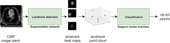
Materials and methods: This study presents a tailor-made neural network architecture for detection of 7 distinctive anatomic landmarks in CMR images of patients with hypoplastic left heart syndrome (HLHS) in Fontan circulation or healthy controls and demonstrates the potential of the spatial arrangement of the landmarks to identify HLHS. The method was applied to the axial SSFP CMR scans of 46 patients with HLHS and 33 healthy controls.
Results: The displacement between predicted and annotated landmark had a standard deviation of 8-17 mm and was larger than the interobserver variability by a factor of 1.1-2.0. A high overall classification accuracy of 98.7% was achieved.
Discussion: Decoupling the identification of clinically meaningful anatomic landmarks from the actual classification improved transparency of classification results. Information from such automated analysis could be used to quickly jump to anatomic positions and guide the physician more efficiently through the analysis depending on the detected condition, which may ultimately improve work flow and save analysis time.
期刊介绍:
MAGMA is a multidisciplinary international journal devoted to the publication of articles on all aspects of magnetic resonance techniques and their applications in medicine and biology. MAGMA currently publishes research papers, reviews, letters to the editor, and commentaries, six times a year. The subject areas covered by MAGMA include:
advances in materials, hardware and software in magnetic resonance technology,
new developments and results in research and practical applications of magnetic resonance imaging and spectroscopy related to biology and medicine,
study of animal models and intact cells using magnetic resonance,
reports of clinical trials on humans and clinical validation of magnetic resonance protocols.

 求助内容:
求助内容: 应助结果提醒方式:


