{"title":"在对怀孕患者进行 CT 暴露时,比较胎儿模型和女性模型中的代用器官。","authors":"Mohamed Khaldoun Badawy, Kashish Kashish, Shay Payne, Maeve Masterson","doi":"10.1007/s13246-024-01383-3","DOIUrl":null,"url":null,"abstract":"<p><p>With the rising use of Computed Tomography (CT) in diagnostic radiology, there are concerns regarding radiation exposure to sensitive groups, including pregnant patients. Accurately determining the radiation dose to the fetus during CT scans is essential to balance diagnostic efficacy with patient safety. This study assessed the accuracy of using the female uterus as a surrogate for fetal radiation dose during CT imaging. The study used common CT protocols to encompass various scenarios, including primary beam, scatter, and partial exposure. The computational program NCICT was used to calculate radiation doses for an adult female and a fetus phantom. The study highlighted that using the uterus for dose estimation can result in consistent underestimations of the effective dose, particularly when the fetus lies within the primary radiation beam. These discrepancies may influence clinical decisions, affecting care strategies and perceptions of associated risks. In conclusion, while the female uterus can indicate fetal radiation dose if the fetus is outside the primary beam, it is unreliable when the fetus is within the primary beam. More reliable abdomen/pelvic organs were recommended.</p>","PeriodicalId":48490,"journal":{"name":"Physical and Engineering Sciences in Medicine","volume":null,"pages":null},"PeriodicalIF":2.4000,"publicationDate":"2024-06-01","publicationTypes":"Journal Article","fieldsOfStudy":null,"isOpenAccess":false,"openAccessPdf":"https://www.ncbi.nlm.nih.gov/pmc/articles/PMC11166780/pdf/","citationCount":"0","resultStr":"{\"title\":\"Comparing fetal phantoms with surrogate organs in female phantoms during CT exposure of pregnant patients.\",\"authors\":\"Mohamed Khaldoun Badawy, Kashish Kashish, Shay Payne, Maeve Masterson\",\"doi\":\"10.1007/s13246-024-01383-3\",\"DOIUrl\":null,\"url\":null,\"abstract\":\"<p><p>With the rising use of Computed Tomography (CT) in diagnostic radiology, there are concerns regarding radiation exposure to sensitive groups, including pregnant patients. Accurately determining the radiation dose to the fetus during CT scans is essential to balance diagnostic efficacy with patient safety. This study assessed the accuracy of using the female uterus as a surrogate for fetal radiation dose during CT imaging. The study used common CT protocols to encompass various scenarios, including primary beam, scatter, and partial exposure. The computational program NCICT was used to calculate radiation doses for an adult female and a fetus phantom. The study highlighted that using the uterus for dose estimation can result in consistent underestimations of the effective dose, particularly when the fetus lies within the primary radiation beam. These discrepancies may influence clinical decisions, affecting care strategies and perceptions of associated risks. In conclusion, while the female uterus can indicate fetal radiation dose if the fetus is outside the primary beam, it is unreliable when the fetus is within the primary beam. More reliable abdomen/pelvic organs were recommended.</p>\",\"PeriodicalId\":48490,\"journal\":{\"name\":\"Physical and Engineering Sciences in Medicine\",\"volume\":null,\"pages\":null},\"PeriodicalIF\":2.4000,\"publicationDate\":\"2024-06-01\",\"publicationTypes\":\"Journal Article\",\"fieldsOfStudy\":null,\"isOpenAccess\":false,\"openAccessPdf\":\"https://www.ncbi.nlm.nih.gov/pmc/articles/PMC11166780/pdf/\",\"citationCount\":\"0\",\"resultStr\":null,\"platform\":\"Semanticscholar\",\"paperid\":null,\"PeriodicalName\":\"Physical and Engineering Sciences in Medicine\",\"FirstCategoryId\":\"3\",\"ListUrlMain\":\"https://doi.org/10.1007/s13246-024-01383-3\",\"RegionNum\":4,\"RegionCategory\":\"医学\",\"ArticlePicture\":[],\"TitleCN\":null,\"AbstractTextCN\":null,\"PMCID\":null,\"EPubDate\":\"2024/1/11 0:00:00\",\"PubModel\":\"Epub\",\"JCR\":\"Q3\",\"JCRName\":\"ENGINEERING, BIOMEDICAL\",\"Score\":null,\"Total\":0}","platform":"Semanticscholar","paperid":null,"PeriodicalName":"Physical and Engineering Sciences in Medicine","FirstCategoryId":"3","ListUrlMain":"https://doi.org/10.1007/s13246-024-01383-3","RegionNum":4,"RegionCategory":"医学","ArticlePicture":[],"TitleCN":null,"AbstractTextCN":null,"PMCID":null,"EPubDate":"2024/1/11 0:00:00","PubModel":"Epub","JCR":"Q3","JCRName":"ENGINEERING, BIOMEDICAL","Score":null,"Total":0}
Comparing fetal phantoms with surrogate organs in female phantoms during CT exposure of pregnant patients.
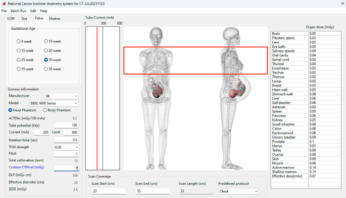
With the rising use of Computed Tomography (CT) in diagnostic radiology, there are concerns regarding radiation exposure to sensitive groups, including pregnant patients. Accurately determining the radiation dose to the fetus during CT scans is essential to balance diagnostic efficacy with patient safety. This study assessed the accuracy of using the female uterus as a surrogate for fetal radiation dose during CT imaging. The study used common CT protocols to encompass various scenarios, including primary beam, scatter, and partial exposure. The computational program NCICT was used to calculate radiation doses for an adult female and a fetus phantom. The study highlighted that using the uterus for dose estimation can result in consistent underestimations of the effective dose, particularly when the fetus lies within the primary radiation beam. These discrepancies may influence clinical decisions, affecting care strategies and perceptions of associated risks. In conclusion, while the female uterus can indicate fetal radiation dose if the fetus is outside the primary beam, it is unreliable when the fetus is within the primary beam. More reliable abdomen/pelvic organs were recommended.
 求助内容:
求助内容: 应助结果提醒方式:


