Adriana Grigoraş, Cornelia Amălinei, Irina Draga Căruntu, Constantin Cristian Grigoraş, Irina Rodica Chiseliţă, Radu Adrian Crişan-Dabija
{"title":"有症状的心包囊肿及其诊断难题。","authors":"Adriana Grigoraş, Cornelia Amălinei, Irina Draga Căruntu, Constantin Cristian Grigoraş, Irina Rodica Chiseliţă, Radu Adrian Crişan-Dabija","doi":"10.47162/RJME.64.4.08","DOIUrl":null,"url":null,"abstract":"<p><p>Pericardial cysts (PCs) or pleuropericardial cysts are rare congenital mediastinal lesions with an approximate incidence of one in 100 000 persons. Usually, they are asymptomatic, being incidentally discovered during a routine chest imaging examination or an autopsy exam. The study involved a retrospective evaluation of clinicopathological findings in a 6-year series of PCs, treated in the Clinic of Pulmonary Diseases, Iaşi, Romania. A group of five cases of PCs, four females and one male, were evaluated. All patients displayed different symptoms, such as dyspnea, chest pain, chronic cough, fatigue, palpitation, and epigastric pain. The cystic lesions were located in the right and left cardiophrenic angle, in four cases, and in the central mediastinum in a single case. The lesions had a fluid content and a maximum diameter that ranged between 35 and 95 mm. The microscopic examination of the surgical resection tissues revealed a thin connective tissue wall without any associated smooth muscle cells. The loose connective tissue band was lined by a layer of mesothelial cells with no cellular atypia, which displayed discrete papillary projections, in one case. Although PCs are rare incidental findings, they should be considered in differential diagnoses of mediastinal cysts, especially as they are associated with non-specific symptoms. Furthermore, considering the possibility of development of severe complications, PCs should be thoroughly explored for suitable patients' management.</p>","PeriodicalId":54447,"journal":{"name":"Romanian Journal of Morphology and Embryology","volume":"64 4","pages":"517-525"},"PeriodicalIF":1.5000,"publicationDate":"2023-10-01","publicationTypes":"Journal Article","fieldsOfStudy":null,"isOpenAccess":false,"openAccessPdf":"https://www.ncbi.nlm.nih.gov/pmc/articles/PMC10863696/pdf/","citationCount":"0","resultStr":"{\"title\":\"Symptomatic pericardial cysts and dilemmas in their diagnosis.\",\"authors\":\"Adriana Grigoraş, Cornelia Amălinei, Irina Draga Căruntu, Constantin Cristian Grigoraş, Irina Rodica Chiseliţă, Radu Adrian Crişan-Dabija\",\"doi\":\"10.47162/RJME.64.4.08\",\"DOIUrl\":null,\"url\":null,\"abstract\":\"<p><p>Pericardial cysts (PCs) or pleuropericardial cysts are rare congenital mediastinal lesions with an approximate incidence of one in 100 000 persons. Usually, they are asymptomatic, being incidentally discovered during a routine chest imaging examination or an autopsy exam. The study involved a retrospective evaluation of clinicopathological findings in a 6-year series of PCs, treated in the Clinic of Pulmonary Diseases, Iaşi, Romania. A group of five cases of PCs, four females and one male, were evaluated. All patients displayed different symptoms, such as dyspnea, chest pain, chronic cough, fatigue, palpitation, and epigastric pain. The cystic lesions were located in the right and left cardiophrenic angle, in four cases, and in the central mediastinum in a single case. The lesions had a fluid content and a maximum diameter that ranged between 35 and 95 mm. The microscopic examination of the surgical resection tissues revealed a thin connective tissue wall without any associated smooth muscle cells. The loose connective tissue band was lined by a layer of mesothelial cells with no cellular atypia, which displayed discrete papillary projections, in one case. Although PCs are rare incidental findings, they should be considered in differential diagnoses of mediastinal cysts, especially as they are associated with non-specific symptoms. Furthermore, considering the possibility of development of severe complications, PCs should be thoroughly explored for suitable patients' management.</p>\",\"PeriodicalId\":54447,\"journal\":{\"name\":\"Romanian Journal of Morphology and Embryology\",\"volume\":\"64 4\",\"pages\":\"517-525\"},\"PeriodicalIF\":1.5000,\"publicationDate\":\"2023-10-01\",\"publicationTypes\":\"Journal Article\",\"fieldsOfStudy\":null,\"isOpenAccess\":false,\"openAccessPdf\":\"https://www.ncbi.nlm.nih.gov/pmc/articles/PMC10863696/pdf/\",\"citationCount\":\"0\",\"resultStr\":null,\"platform\":\"Semanticscholar\",\"paperid\":null,\"PeriodicalName\":\"Romanian Journal of Morphology and Embryology\",\"FirstCategoryId\":\"3\",\"ListUrlMain\":\"https://doi.org/10.47162/RJME.64.4.08\",\"RegionNum\":4,\"RegionCategory\":\"医学\",\"ArticlePicture\":[],\"TitleCN\":null,\"AbstractTextCN\":null,\"PMCID\":null,\"EPubDate\":\"\",\"PubModel\":\"\",\"JCR\":\"Q4\",\"JCRName\":\"DEVELOPMENTAL BIOLOGY\",\"Score\":null,\"Total\":0}","platform":"Semanticscholar","paperid":null,"PeriodicalName":"Romanian Journal of Morphology and Embryology","FirstCategoryId":"3","ListUrlMain":"https://doi.org/10.47162/RJME.64.4.08","RegionNum":4,"RegionCategory":"医学","ArticlePicture":[],"TitleCN":null,"AbstractTextCN":null,"PMCID":null,"EPubDate":"","PubModel":"","JCR":"Q4","JCRName":"DEVELOPMENTAL BIOLOGY","Score":null,"Total":0}
Symptomatic pericardial cysts and dilemmas in their diagnosis.
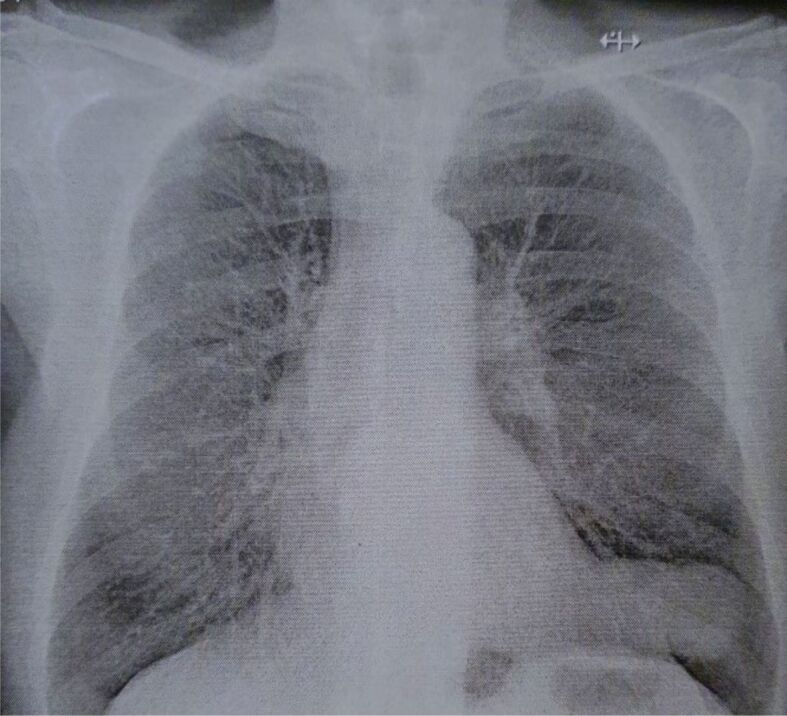
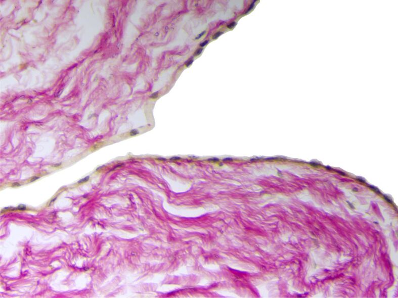
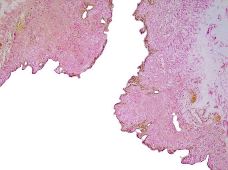
Pericardial cysts (PCs) or pleuropericardial cysts are rare congenital mediastinal lesions with an approximate incidence of one in 100 000 persons. Usually, they are asymptomatic, being incidentally discovered during a routine chest imaging examination or an autopsy exam. The study involved a retrospective evaluation of clinicopathological findings in a 6-year series of PCs, treated in the Clinic of Pulmonary Diseases, Iaşi, Romania. A group of five cases of PCs, four females and one male, were evaluated. All patients displayed different symptoms, such as dyspnea, chest pain, chronic cough, fatigue, palpitation, and epigastric pain. The cystic lesions were located in the right and left cardiophrenic angle, in four cases, and in the central mediastinum in a single case. The lesions had a fluid content and a maximum diameter that ranged between 35 and 95 mm. The microscopic examination of the surgical resection tissues revealed a thin connective tissue wall without any associated smooth muscle cells. The loose connective tissue band was lined by a layer of mesothelial cells with no cellular atypia, which displayed discrete papillary projections, in one case. Although PCs are rare incidental findings, they should be considered in differential diagnoses of mediastinal cysts, especially as they are associated with non-specific symptoms. Furthermore, considering the possibility of development of severe complications, PCs should be thoroughly explored for suitable patients' management.
期刊介绍:
Romanian Journal of Morphology and Embryology (Rom J Morphol Embryol) publishes studies on all aspects of normal morphology and human comparative and experimental pathology. The Journal accepts only researches that utilize modern investigation methods (studies of anatomy, pathology, cytopathology, immunohistochemistry, histochemistry, immunology, morphometry, molecular and cellular biology, electronic microscopy, etc.).
 求助内容:
求助内容: 应助结果提醒方式:


