{"title":"STING 上调通过上调 TBK1 和激活 NF-κB 信号通路介导狼疮肾炎中的铁蛋白沉积和炎症反应","authors":"Jinshu Chen, Pihou Chen, Yijin Song, Jiaxin Wei, Fan Wu, Jing Sun, Zhiquan Xu","doi":"10.1007/s12038-023-00381-z","DOIUrl":null,"url":null,"abstract":"<p>Accumulated evidence implicates lipid peroxidation as a key mechanism contributing to the pathogenesis of lupus nephritis (LN). Ferroptosis is a specialized form of cell death induced by loss or deficient activity of the glutathione peroxidase 4 (GPX4) and decreased clearance of polyunsaturated fatty acid hydroperoxides. STING production may lead to the occurrence of intracellular lipid peroxidation, ultimately triggering ferroptosis, but it has not been clarified whether STING can aggravate LN via ferroptosis. The adjacent normal kidney tissues from renal cell carcinoma and biopsied kidney tissue samples from LN patients were used for research, and the expression of STING protein in kidney tissue was detected by immunohistochemistry and RT-qPCR. MRL/lpr mice, a model of LN, were used to detect STING expression in kidney tissue. STING expression in the kidney tissue of MRL/lpr mice was knocked down by sh-STING-AAV, and then levels of 4-HNE, MDA, ROS, iron ion, blood urea nitrogen and serum creatinine, IL-6, IL-1β, and TNF-α, and the protein expression of STING, <i>TBK1</i>, NF-κB, GPX4, ACSL4, and SLC7A11 were subsequently examined. STING was elevated in the kidney tissue of LN patients and MRL/lpr mice. Compared with the MRL/lpr group, liproxstatin-1 or ferrostatin-1 treatment alleviated ferroptosis-related indicators 4-HNE, MDA, ROS, iron ion release, and GPX4 and SLC7A1 expression, whereas the treatment enhanced ACSL4 expression. STING interference observably decreased 4-HNE, ROS, MDA, iron ion, STING, and ACSL4 levels, and increased GPX4 and SLC7A11 expression in MRL/lpr mice kidney tissues. Besides, inhibition of STING reduced kidney tissue damage and inflammatory cell infiltration in MRL/lpr mice, and levels of serum creatinine, blood urea nitrogen, serum anti-double-stranded DNA antibody, inflammatory factors IL-6, IL-1β, and TNF-α, as well as phosphorylation of NF-κB were all significantly decreased in MRL/lpr mice. <i>TBK1</i> overexpression reversed the impact of STING inhibition on ferroptosis and inflammatory response. STING contributed to ferroptosis and inflammatory response by activating the <i>TBK1</i>/NF-κB pathway, suggesting that STING may be a potent therapeutic target in LN.</p>","PeriodicalId":15171,"journal":{"name":"Journal of Biosciences","volume":"45 6 1","pages":""},"PeriodicalIF":2.1000,"publicationDate":"2024-01-02","publicationTypes":"Journal Article","fieldsOfStudy":null,"isOpenAccess":false,"openAccessPdf":"","citationCount":"0","resultStr":"{\"title\":\"STING upregulation mediates ferroptosis and inflammatory response in lupus nephritis by upregulating TBK1 and activating NF-κB signal pathway\",\"authors\":\"Jinshu Chen, Pihou Chen, Yijin Song, Jiaxin Wei, Fan Wu, Jing Sun, Zhiquan Xu\",\"doi\":\"10.1007/s12038-023-00381-z\",\"DOIUrl\":null,\"url\":null,\"abstract\":\"<p>Accumulated evidence implicates lipid peroxidation as a key mechanism contributing to the pathogenesis of lupus nephritis (LN). Ferroptosis is a specialized form of cell death induced by loss or deficient activity of the glutathione peroxidase 4 (GPX4) and decreased clearance of polyunsaturated fatty acid hydroperoxides. STING production may lead to the occurrence of intracellular lipid peroxidation, ultimately triggering ferroptosis, but it has not been clarified whether STING can aggravate LN via ferroptosis. The adjacent normal kidney tissues from renal cell carcinoma and biopsied kidney tissue samples from LN patients were used for research, and the expression of STING protein in kidney tissue was detected by immunohistochemistry and RT-qPCR. MRL/lpr mice, a model of LN, were used to detect STING expression in kidney tissue. STING expression in the kidney tissue of MRL/lpr mice was knocked down by sh-STING-AAV, and then levels of 4-HNE, MDA, ROS, iron ion, blood urea nitrogen and serum creatinine, IL-6, IL-1β, and TNF-α, and the protein expression of STING, <i>TBK1</i>, NF-κB, GPX4, ACSL4, and SLC7A11 were subsequently examined. STING was elevated in the kidney tissue of LN patients and MRL/lpr mice. Compared with the MRL/lpr group, liproxstatin-1 or ferrostatin-1 treatment alleviated ferroptosis-related indicators 4-HNE, MDA, ROS, iron ion release, and GPX4 and SLC7A1 expression, whereas the treatment enhanced ACSL4 expression. STING interference observably decreased 4-HNE, ROS, MDA, iron ion, STING, and ACSL4 levels, and increased GPX4 and SLC7A11 expression in MRL/lpr mice kidney tissues. Besides, inhibition of STING reduced kidney tissue damage and inflammatory cell infiltration in MRL/lpr mice, and levels of serum creatinine, blood urea nitrogen, serum anti-double-stranded DNA antibody, inflammatory factors IL-6, IL-1β, and TNF-α, as well as phosphorylation of NF-κB were all significantly decreased in MRL/lpr mice. <i>TBK1</i> overexpression reversed the impact of STING inhibition on ferroptosis and inflammatory response. STING contributed to ferroptosis and inflammatory response by activating the <i>TBK1</i>/NF-κB pathway, suggesting that STING may be a potent therapeutic target in LN.</p>\",\"PeriodicalId\":15171,\"journal\":{\"name\":\"Journal of Biosciences\",\"volume\":\"45 6 1\",\"pages\":\"\"},\"PeriodicalIF\":2.1000,\"publicationDate\":\"2024-01-02\",\"publicationTypes\":\"Journal Article\",\"fieldsOfStudy\":null,\"isOpenAccess\":false,\"openAccessPdf\":\"\",\"citationCount\":\"0\",\"resultStr\":null,\"platform\":\"Semanticscholar\",\"paperid\":null,\"PeriodicalName\":\"Journal of Biosciences\",\"FirstCategoryId\":\"99\",\"ListUrlMain\":\"https://doi.org/10.1007/s12038-023-00381-z\",\"RegionNum\":4,\"RegionCategory\":\"生物学\",\"ArticlePicture\":[],\"TitleCN\":null,\"AbstractTextCN\":null,\"PMCID\":null,\"EPubDate\":\"\",\"PubModel\":\"\",\"JCR\":\"Q2\",\"JCRName\":\"BIOLOGY\",\"Score\":null,\"Total\":0}","platform":"Semanticscholar","paperid":null,"PeriodicalName":"Journal of Biosciences","FirstCategoryId":"99","ListUrlMain":"https://doi.org/10.1007/s12038-023-00381-z","RegionNum":4,"RegionCategory":"生物学","ArticlePicture":[],"TitleCN":null,"AbstractTextCN":null,"PMCID":null,"EPubDate":"","PubModel":"","JCR":"Q2","JCRName":"BIOLOGY","Score":null,"Total":0}
STING upregulation mediates ferroptosis and inflammatory response in lupus nephritis by upregulating TBK1 and activating NF-κB signal pathway
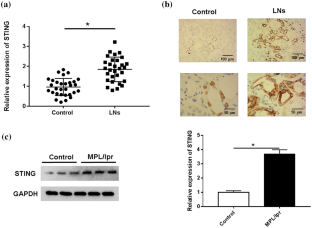
Accumulated evidence implicates lipid peroxidation as a key mechanism contributing to the pathogenesis of lupus nephritis (LN). Ferroptosis is a specialized form of cell death induced by loss or deficient activity of the glutathione peroxidase 4 (GPX4) and decreased clearance of polyunsaturated fatty acid hydroperoxides. STING production may lead to the occurrence of intracellular lipid peroxidation, ultimately triggering ferroptosis, but it has not been clarified whether STING can aggravate LN via ferroptosis. The adjacent normal kidney tissues from renal cell carcinoma and biopsied kidney tissue samples from LN patients were used for research, and the expression of STING protein in kidney tissue was detected by immunohistochemistry and RT-qPCR. MRL/lpr mice, a model of LN, were used to detect STING expression in kidney tissue. STING expression in the kidney tissue of MRL/lpr mice was knocked down by sh-STING-AAV, and then levels of 4-HNE, MDA, ROS, iron ion, blood urea nitrogen and serum creatinine, IL-6, IL-1β, and TNF-α, and the protein expression of STING, TBK1, NF-κB, GPX4, ACSL4, and SLC7A11 were subsequently examined. STING was elevated in the kidney tissue of LN patients and MRL/lpr mice. Compared with the MRL/lpr group, liproxstatin-1 or ferrostatin-1 treatment alleviated ferroptosis-related indicators 4-HNE, MDA, ROS, iron ion release, and GPX4 and SLC7A1 expression, whereas the treatment enhanced ACSL4 expression. STING interference observably decreased 4-HNE, ROS, MDA, iron ion, STING, and ACSL4 levels, and increased GPX4 and SLC7A11 expression in MRL/lpr mice kidney tissues. Besides, inhibition of STING reduced kidney tissue damage and inflammatory cell infiltration in MRL/lpr mice, and levels of serum creatinine, blood urea nitrogen, serum anti-double-stranded DNA antibody, inflammatory factors IL-6, IL-1β, and TNF-α, as well as phosphorylation of NF-κB were all significantly decreased in MRL/lpr mice. TBK1 overexpression reversed the impact of STING inhibition on ferroptosis and inflammatory response. STING contributed to ferroptosis and inflammatory response by activating the TBK1/NF-κB pathway, suggesting that STING may be a potent therapeutic target in LN.
期刊介绍:
The Journal of Biosciences is a quarterly journal published by the Indian Academy of Sciences, Bangalore. It covers all areas of Biology and is the premier journal in the country within its scope. It is indexed in Current Contents and other standard Biological and Medical databases. The Journal of Biosciences began in 1934 as the Proceedings of the Indian Academy of Sciences (Section B). This continued until 1978 when it was split into three parts : Proceedings-Animal Sciences, Proceedings-Plant Sciences and Proceedings-Experimental Biology. Proceedings-Experimental Biology was renamed Journal of Biosciences in 1979; and in 1991, Proceedings-Animal Sciences and Proceedings-Plant Sciences merged with it.
 求助内容:
求助内容: 应助结果提醒方式:


