Bogun Jang, Hyesung Kim, Su-Hyung Lee, Yoonkyung Won, Izumi Kaji, Robert J Coffey, Eunyoung Choi, James R Goldenring
{"title":"胃化生和发育不良过程中的动态簇细胞扩增。","authors":"Bogun Jang, Hyesung Kim, Su-Hyung Lee, Yoonkyung Won, Izumi Kaji, Robert J Coffey, Eunyoung Choi, James R Goldenring","doi":"10.1002/cjp2.352","DOIUrl":null,"url":null,"abstract":"<p>Tuft cells are chemosensory cells associated with luminal homeostasis, immune response, and tumorigenesis in the gastrointestinal tract. We aimed to elucidate alterations in tuft cell populations during gastric atrophy and tumorigenesis in humans with correlative comparison to relevant mouse models. Tuft cell distribution was determined in human stomachs from organ donors and in gastric pathologies including Ménétrier's disease, <i>Helicobacter pylori</i> gastritis, intestinal metaplasia (IM), and gastric tumors. Tuft cell populations were examined in <i>Lrig1-Kras</i><sup><i>G12D</i></sup>, <i>Mist1-Kras</i><sup><i>G12D</i></sup>, and MT-TGFα mice. Tuft cells were evenly distributed throughout the entire normal human stomach, primarily concentrated in the isthmal region in the fundus. Ménétrier's disease stomach showed increased tuft cells. Similarly, Lrig1-Kras mice and mice overexpressing TGFα showed marked foveolar hyperplasia and expanded tuft cell populations. Human stomach with IM or dysplasia also showed increased tuft cell numbers. Similarly, Mist1-Kras mice had increased numbers of tuft cells during metaplasia and dysplasia development. In human gastric cancers, tuft cells were rarely observed, but showed positive associations with well-differentiated lesions. In mouse gastric cancer xenografts, tuft cells were restricted to dysplastic well-differentiated mucinous cysts and were lost in less differentiated cancers. Taken together, tuft cell populations increased in atrophic human gastric pathologies, metaplasia, and dysplasia, but were decreased in gastric cancers. Similar findings were observed in mouse models, suggesting that, while tuft cells are associated with precancerous pathologies, their loss is most associated with the progression to invasive cancer.</p>","PeriodicalId":48612,"journal":{"name":"Journal of Pathology Clinical Research","volume":"10 1","pages":""},"PeriodicalIF":3.4000,"publicationDate":"2023-12-20","publicationTypes":"Journal Article","fieldsOfStudy":null,"isOpenAccess":false,"openAccessPdf":"https://onlinelibrary.wiley.com/doi/epdf/10.1002/cjp2.352","citationCount":"0","resultStr":"{\"title\":\"Dynamic tuft cell expansion during gastric metaplasia and dysplasia\",\"authors\":\"Bogun Jang, Hyesung Kim, Su-Hyung Lee, Yoonkyung Won, Izumi Kaji, Robert J Coffey, Eunyoung Choi, James R Goldenring\",\"doi\":\"10.1002/cjp2.352\",\"DOIUrl\":null,\"url\":null,\"abstract\":\"<p>Tuft cells are chemosensory cells associated with luminal homeostasis, immune response, and tumorigenesis in the gastrointestinal tract. We aimed to elucidate alterations in tuft cell populations during gastric atrophy and tumorigenesis in humans with correlative comparison to relevant mouse models. Tuft cell distribution was determined in human stomachs from organ donors and in gastric pathologies including Ménétrier's disease, <i>Helicobacter pylori</i> gastritis, intestinal metaplasia (IM), and gastric tumors. Tuft cell populations were examined in <i>Lrig1-Kras</i><sup><i>G12D</i></sup>, <i>Mist1-Kras</i><sup><i>G12D</i></sup>, and MT-TGFα mice. Tuft cells were evenly distributed throughout the entire normal human stomach, primarily concentrated in the isthmal region in the fundus. Ménétrier's disease stomach showed increased tuft cells. Similarly, Lrig1-Kras mice and mice overexpressing TGFα showed marked foveolar hyperplasia and expanded tuft cell populations. Human stomach with IM or dysplasia also showed increased tuft cell numbers. Similarly, Mist1-Kras mice had increased numbers of tuft cells during metaplasia and dysplasia development. In human gastric cancers, tuft cells were rarely observed, but showed positive associations with well-differentiated lesions. In mouse gastric cancer xenografts, tuft cells were restricted to dysplastic well-differentiated mucinous cysts and were lost in less differentiated cancers. Taken together, tuft cell populations increased in atrophic human gastric pathologies, metaplasia, and dysplasia, but were decreased in gastric cancers. Similar findings were observed in mouse models, suggesting that, while tuft cells are associated with precancerous pathologies, their loss is most associated with the progression to invasive cancer.</p>\",\"PeriodicalId\":48612,\"journal\":{\"name\":\"Journal of Pathology Clinical Research\",\"volume\":\"10 1\",\"pages\":\"\"},\"PeriodicalIF\":3.4000,\"publicationDate\":\"2023-12-20\",\"publicationTypes\":\"Journal Article\",\"fieldsOfStudy\":null,\"isOpenAccess\":false,\"openAccessPdf\":\"https://onlinelibrary.wiley.com/doi/epdf/10.1002/cjp2.352\",\"citationCount\":\"0\",\"resultStr\":null,\"platform\":\"Semanticscholar\",\"paperid\":null,\"PeriodicalName\":\"Journal of Pathology Clinical Research\",\"FirstCategoryId\":\"3\",\"ListUrlMain\":\"https://onlinelibrary.wiley.com/doi/10.1002/cjp2.352\",\"RegionNum\":2,\"RegionCategory\":\"医学\",\"ArticlePicture\":[],\"TitleCN\":null,\"AbstractTextCN\":null,\"PMCID\":null,\"EPubDate\":\"\",\"PubModel\":\"\",\"JCR\":\"Q1\",\"JCRName\":\"PATHOLOGY\",\"Score\":null,\"Total\":0}","platform":"Semanticscholar","paperid":null,"PeriodicalName":"Journal of Pathology Clinical Research","FirstCategoryId":"3","ListUrlMain":"https://onlinelibrary.wiley.com/doi/10.1002/cjp2.352","RegionNum":2,"RegionCategory":"医学","ArticlePicture":[],"TitleCN":null,"AbstractTextCN":null,"PMCID":null,"EPubDate":"","PubModel":"","JCR":"Q1","JCRName":"PATHOLOGY","Score":null,"Total":0}
Dynamic tuft cell expansion during gastric metaplasia and dysplasia
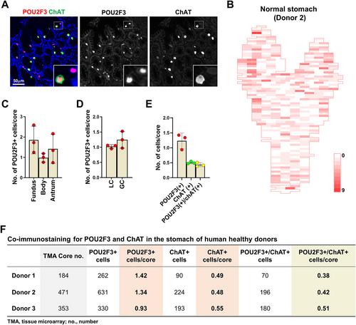
Tuft cells are chemosensory cells associated with luminal homeostasis, immune response, and tumorigenesis in the gastrointestinal tract. We aimed to elucidate alterations in tuft cell populations during gastric atrophy and tumorigenesis in humans with correlative comparison to relevant mouse models. Tuft cell distribution was determined in human stomachs from organ donors and in gastric pathologies including Ménétrier's disease, Helicobacter pylori gastritis, intestinal metaplasia (IM), and gastric tumors. Tuft cell populations were examined in Lrig1-KrasG12D, Mist1-KrasG12D, and MT-TGFα mice. Tuft cells were evenly distributed throughout the entire normal human stomach, primarily concentrated in the isthmal region in the fundus. Ménétrier's disease stomach showed increased tuft cells. Similarly, Lrig1-Kras mice and mice overexpressing TGFα showed marked foveolar hyperplasia and expanded tuft cell populations. Human stomach with IM or dysplasia also showed increased tuft cell numbers. Similarly, Mist1-Kras mice had increased numbers of tuft cells during metaplasia and dysplasia development. In human gastric cancers, tuft cells were rarely observed, but showed positive associations with well-differentiated lesions. In mouse gastric cancer xenografts, tuft cells were restricted to dysplastic well-differentiated mucinous cysts and were lost in less differentiated cancers. Taken together, tuft cell populations increased in atrophic human gastric pathologies, metaplasia, and dysplasia, but were decreased in gastric cancers. Similar findings were observed in mouse models, suggesting that, while tuft cells are associated with precancerous pathologies, their loss is most associated with the progression to invasive cancer.
期刊介绍:
The Journal of Pathology: Clinical Research and The Journal of Pathology serve as translational bridges between basic biomedical science and clinical medicine with particular emphasis on, but not restricted to, tissue based studies.
The focus of The Journal of Pathology: Clinical Research is the publication of studies that illuminate the clinical relevance of research in the broad area of the study of disease. Appropriately powered and validated studies with novel diagnostic, prognostic and predictive significance, and biomarker discover and validation, will be welcomed. Studies with a predominantly mechanistic basis will be more appropriate for the companion Journal of Pathology.
 求助内容:
求助内容: 应助结果提醒方式:


