{"title":"血吸虫病与结直肠癌的临床、影像和病理特征的联合分析。","authors":"Fang Zhang, XiaoShuang Wang, YuanTing Zhu, Peng Xia","doi":"10.3389/pore.2023.1611396","DOIUrl":null,"url":null,"abstract":"<p><p>This study aims to examine and compare clinical, radiological, and pathological data between colorectal cancer (CRC) patients with and without schistosomiasis and uncover distinctive CRC characteristics when accompanied by schistosomiasis. This retrospective study is based on data collected from 341 patients diagnosed with CRC post-surgery and pathology. Of these patients, 101 (Group A) were diagnosed with colorectal cancer co-occurring with schistosomiasis (CRC-S), while 240 patients (Group B) were diagnosed with colorectal cancer without concurrent schistosomiasis (CRC-NS). Both groups were compared and analyzed based on their clinical data, imaging-based TNM staging, lymph node metastasis, nerve invasion, vascular cancer thrombus, and histopathological differentiation. A Chi-squared test revealed a significant difference in gender distribution between the patients with CRC-S (Group A) and CRC-NS (Group B), with a <i>p</i> -value of 0.043 and χ<sup>2</sup> = 4.115. Specifically, a higher incidence rate was observed among males in Group A. There was a difference in the overall distribution of TNM staging between the two groups (<i>p</i> = 0.034, χ<sup>2</sup> = 6.764). After pairwise comparison, a statistically significant difference was observed in the T3 stage (<i>p</i> <0.05). The proportion of the T3 stage in Group A was significantly higher than that in Group B, indicating certain advantages. There was a difference in postoperative histopathological grading between the two groups (<i>p</i> = 0.005, χ<sup>2</sup> = 10.626). After pairwise comparison, a statistically significant difference was observed between the well-differentiated adenocarcinoma and the moderately and poorly differentiated adenocarcinoma (<i>p</i> <0.05), with a higher proportion of welldifferentiated patients in Group A compared to Group B. There was no significant difference in age, lymph node metastasis, nerve invasion, and vascular invasion between the two groups of patients (<i>p</i> > 0.05). Among the 101 patients with CRC-S, 87 (86%) showed linear calcification on CT imaging. Patients with CRC-S are mainly male, with tumor staging mostly in the middle stage, high tumor differentiation, and low malignancy. CT imaging can help identify the presence of lumps and linear calcification indicative of schistosome deposits. MRI can early clarify TNM staging and determine the presence of lymph node metastasis and nerve and vascular invasion.</p>","PeriodicalId":19981,"journal":{"name":"Pathology & Oncology Research","volume":"29 ","pages":"1611396"},"PeriodicalIF":2.3000,"publicationDate":"2023-11-30","publicationTypes":"Journal Article","fieldsOfStudy":null,"isOpenAccess":false,"openAccessPdf":"https://www.ncbi.nlm.nih.gov/pmc/articles/PMC10719402/pdf/","citationCount":"0","resultStr":"{\"title\":\"Conjoint analysis of clinical, imaging, and pathological features of schistosomiasis and colorectal cancer.\",\"authors\":\"Fang Zhang, XiaoShuang Wang, YuanTing Zhu, Peng Xia\",\"doi\":\"10.3389/pore.2023.1611396\",\"DOIUrl\":null,\"url\":null,\"abstract\":\"<p><p>This study aims to examine and compare clinical, radiological, and pathological data between colorectal cancer (CRC) patients with and without schistosomiasis and uncover distinctive CRC characteristics when accompanied by schistosomiasis. This retrospective study is based on data collected from 341 patients diagnosed with CRC post-surgery and pathology. Of these patients, 101 (Group A) were diagnosed with colorectal cancer co-occurring with schistosomiasis (CRC-S), while 240 patients (Group B) were diagnosed with colorectal cancer without concurrent schistosomiasis (CRC-NS). Both groups were compared and analyzed based on their clinical data, imaging-based TNM staging, lymph node metastasis, nerve invasion, vascular cancer thrombus, and histopathological differentiation. A Chi-squared test revealed a significant difference in gender distribution between the patients with CRC-S (Group A) and CRC-NS (Group B), with a <i>p</i> -value of 0.043 and χ<sup>2</sup> = 4.115. Specifically, a higher incidence rate was observed among males in Group A. There was a difference in the overall distribution of TNM staging between the two groups (<i>p</i> = 0.034, χ<sup>2</sup> = 6.764). After pairwise comparison, a statistically significant difference was observed in the T3 stage (<i>p</i> <0.05). The proportion of the T3 stage in Group A was significantly higher than that in Group B, indicating certain advantages. There was a difference in postoperative histopathological grading between the two groups (<i>p</i> = 0.005, χ<sup>2</sup> = 10.626). After pairwise comparison, a statistically significant difference was observed between the well-differentiated adenocarcinoma and the moderately and poorly differentiated adenocarcinoma (<i>p</i> <0.05), with a higher proportion of welldifferentiated patients in Group A compared to Group B. There was no significant difference in age, lymph node metastasis, nerve invasion, and vascular invasion between the two groups of patients (<i>p</i> > 0.05). Among the 101 patients with CRC-S, 87 (86%) showed linear calcification on CT imaging. Patients with CRC-S are mainly male, with tumor staging mostly in the middle stage, high tumor differentiation, and low malignancy. CT imaging can help identify the presence of lumps and linear calcification indicative of schistosome deposits. MRI can early clarify TNM staging and determine the presence of lymph node metastasis and nerve and vascular invasion.</p>\",\"PeriodicalId\":19981,\"journal\":{\"name\":\"Pathology & Oncology Research\",\"volume\":\"29 \",\"pages\":\"1611396\"},\"PeriodicalIF\":2.3000,\"publicationDate\":\"2023-11-30\",\"publicationTypes\":\"Journal Article\",\"fieldsOfStudy\":null,\"isOpenAccess\":false,\"openAccessPdf\":\"https://www.ncbi.nlm.nih.gov/pmc/articles/PMC10719402/pdf/\",\"citationCount\":\"0\",\"resultStr\":null,\"platform\":\"Semanticscholar\",\"paperid\":null,\"PeriodicalName\":\"Pathology & Oncology Research\",\"FirstCategoryId\":\"3\",\"ListUrlMain\":\"https://doi.org/10.3389/pore.2023.1611396\",\"RegionNum\":4,\"RegionCategory\":\"医学\",\"ArticlePicture\":[],\"TitleCN\":null,\"AbstractTextCN\":null,\"PMCID\":null,\"EPubDate\":\"2023/1/1 0:00:00\",\"PubModel\":\"eCollection\",\"JCR\":\"Q3\",\"JCRName\":\"ONCOLOGY\",\"Score\":null,\"Total\":0}","platform":"Semanticscholar","paperid":null,"PeriodicalName":"Pathology & Oncology Research","FirstCategoryId":"3","ListUrlMain":"https://doi.org/10.3389/pore.2023.1611396","RegionNum":4,"RegionCategory":"医学","ArticlePicture":[],"TitleCN":null,"AbstractTextCN":null,"PMCID":null,"EPubDate":"2023/1/1 0:00:00","PubModel":"eCollection","JCR":"Q3","JCRName":"ONCOLOGY","Score":null,"Total":0}
Conjoint analysis of clinical, imaging, and pathological features of schistosomiasis and colorectal cancer.
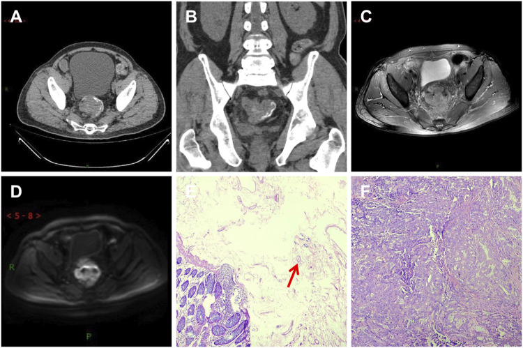
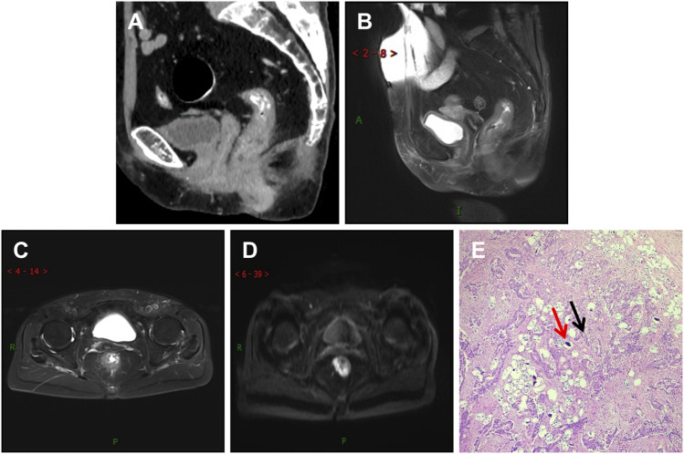
This study aims to examine and compare clinical, radiological, and pathological data between colorectal cancer (CRC) patients with and without schistosomiasis and uncover distinctive CRC characteristics when accompanied by schistosomiasis. This retrospective study is based on data collected from 341 patients diagnosed with CRC post-surgery and pathology. Of these patients, 101 (Group A) were diagnosed with colorectal cancer co-occurring with schistosomiasis (CRC-S), while 240 patients (Group B) were diagnosed with colorectal cancer without concurrent schistosomiasis (CRC-NS). Both groups were compared and analyzed based on their clinical data, imaging-based TNM staging, lymph node metastasis, nerve invasion, vascular cancer thrombus, and histopathological differentiation. A Chi-squared test revealed a significant difference in gender distribution between the patients with CRC-S (Group A) and CRC-NS (Group B), with a p -value of 0.043 and χ2 = 4.115. Specifically, a higher incidence rate was observed among males in Group A. There was a difference in the overall distribution of TNM staging between the two groups (p = 0.034, χ2 = 6.764). After pairwise comparison, a statistically significant difference was observed in the T3 stage (p <0.05). The proportion of the T3 stage in Group A was significantly higher than that in Group B, indicating certain advantages. There was a difference in postoperative histopathological grading between the two groups (p = 0.005, χ2 = 10.626). After pairwise comparison, a statistically significant difference was observed between the well-differentiated adenocarcinoma and the moderately and poorly differentiated adenocarcinoma (p <0.05), with a higher proportion of welldifferentiated patients in Group A compared to Group B. There was no significant difference in age, lymph node metastasis, nerve invasion, and vascular invasion between the two groups of patients (p > 0.05). Among the 101 patients with CRC-S, 87 (86%) showed linear calcification on CT imaging. Patients with CRC-S are mainly male, with tumor staging mostly in the middle stage, high tumor differentiation, and low malignancy. CT imaging can help identify the presence of lumps and linear calcification indicative of schistosome deposits. MRI can early clarify TNM staging and determine the presence of lymph node metastasis and nerve and vascular invasion.
期刊介绍:
Pathology & Oncology Research (POR) is an interdisciplinary Journal at the interface of pathology and oncology including the preclinical and translational research, diagnostics and therapy. Furthermore, POR is an international forum for the rapid communication of reviews, original research, critical and topical reports with excellence and novelty. Published quarterly, POR is dedicated to keeping scientists informed of developments on the selected biomedical fields bridging the gap between basic research and clinical medicine. It is a special aim for POR to promote pathological and oncological publishing activity of colleagues in the Central and East European region. The journal will be of interest to pathologists, and a broad range of experimental and clinical oncologists, and related experts. POR is supported by an acknowledged international advisory board and the Arányi Fundation for modern pathology.
 求助内容:
求助内容: 应助结果提醒方式:


