{"title":"单侧耳蜗给药后不同 AAV 向量的分布比较","authors":"Shuang Han, Zhijiao Xu, Shengyi Wang, Honghai Tang, Shaowei Hu, Hui Wang, Guofang Guan, Yilai Shu","doi":"10.1038/s41434-023-00431-z","DOIUrl":null,"url":null,"abstract":"The adeno-associated virus (AAV) gene therapy has been widely applied to mouse models for deafness. But, AAVs could transduce non-targeted organs after inner ear delivery due to their low cell-type specificity. This study compares transgene expression and biodistribution of AAV1, AAV2, Anc80L65, AAV9, AAV-PHP.B, and AAV-PHP.eB after round window membrane (RWM) injection in neonatal mice. The highest virus concentration was detected in the injected cochlea. AAV2, Anc80L65, AAV9, AAV-PHP.B, and AAV-PHP.eB transduced both inner hair cells (IHCs) and outer hair cells (OHCs) with high efficiency, while AAV1 transduced IHCs with high efficiency but OHCs with low efficiency. All AAV subtypes finitely transduced contralateral inner ear, brain, heart, and liver compared with the injected cochlea. In most brain regions, the enhanced green fluorescent protein (eGFP) expression of AAV1 and AAV2 was lower than that of other four subtypes. We suggested the cochlear aqueduct might be one of routes for vectors instantaneously infiltrating into the brain from the cochlea through a dye tracking test. In summary, our results provide available data for further investigating the biodistribution of vectors through local inner ear injection and afford a reference for selecting AAV serotypes for gene therapy toward deafness.","PeriodicalId":12699,"journal":{"name":"Gene Therapy","volume":"31 3-4","pages":"154-164"},"PeriodicalIF":4.6000,"publicationDate":"2023-12-14","publicationTypes":"Journal Article","fieldsOfStudy":null,"isOpenAccess":false,"openAccessPdf":"","citationCount":"0","resultStr":"{\"title\":\"Distributional comparison of different AAV vectors after unilateral cochlear administration\",\"authors\":\"Shuang Han, Zhijiao Xu, Shengyi Wang, Honghai Tang, Shaowei Hu, Hui Wang, Guofang Guan, Yilai Shu\",\"doi\":\"10.1038/s41434-023-00431-z\",\"DOIUrl\":null,\"url\":null,\"abstract\":\"The adeno-associated virus (AAV) gene therapy has been widely applied to mouse models for deafness. But, AAVs could transduce non-targeted organs after inner ear delivery due to their low cell-type specificity. This study compares transgene expression and biodistribution of AAV1, AAV2, Anc80L65, AAV9, AAV-PHP.B, and AAV-PHP.eB after round window membrane (RWM) injection in neonatal mice. The highest virus concentration was detected in the injected cochlea. AAV2, Anc80L65, AAV9, AAV-PHP.B, and AAV-PHP.eB transduced both inner hair cells (IHCs) and outer hair cells (OHCs) with high efficiency, while AAV1 transduced IHCs with high efficiency but OHCs with low efficiency. All AAV subtypes finitely transduced contralateral inner ear, brain, heart, and liver compared with the injected cochlea. In most brain regions, the enhanced green fluorescent protein (eGFP) expression of AAV1 and AAV2 was lower than that of other four subtypes. We suggested the cochlear aqueduct might be one of routes for vectors instantaneously infiltrating into the brain from the cochlea through a dye tracking test. In summary, our results provide available data for further investigating the biodistribution of vectors through local inner ear injection and afford a reference for selecting AAV serotypes for gene therapy toward deafness.\",\"PeriodicalId\":12699,\"journal\":{\"name\":\"Gene Therapy\",\"volume\":\"31 3-4\",\"pages\":\"154-164\"},\"PeriodicalIF\":4.6000,\"publicationDate\":\"2023-12-14\",\"publicationTypes\":\"Journal Article\",\"fieldsOfStudy\":null,\"isOpenAccess\":false,\"openAccessPdf\":\"\",\"citationCount\":\"0\",\"resultStr\":null,\"platform\":\"Semanticscholar\",\"paperid\":null,\"PeriodicalName\":\"Gene Therapy\",\"FirstCategoryId\":\"3\",\"ListUrlMain\":\"https://www.nature.com/articles/s41434-023-00431-z\",\"RegionNum\":3,\"RegionCategory\":\"医学\",\"ArticlePicture\":[],\"TitleCN\":null,\"AbstractTextCN\":null,\"PMCID\":null,\"EPubDate\":\"\",\"PubModel\":\"\",\"JCR\":\"Q1\",\"JCRName\":\"BIOCHEMISTRY & MOLECULAR BIOLOGY\",\"Score\":null,\"Total\":0}","platform":"Semanticscholar","paperid":null,"PeriodicalName":"Gene Therapy","FirstCategoryId":"3","ListUrlMain":"https://www.nature.com/articles/s41434-023-00431-z","RegionNum":3,"RegionCategory":"医学","ArticlePicture":[],"TitleCN":null,"AbstractTextCN":null,"PMCID":null,"EPubDate":"","PubModel":"","JCR":"Q1","JCRName":"BIOCHEMISTRY & MOLECULAR BIOLOGY","Score":null,"Total":0}
Distributional comparison of different AAV vectors after unilateral cochlear administration
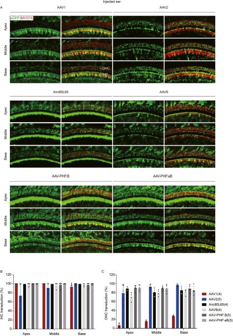
The adeno-associated virus (AAV) gene therapy has been widely applied to mouse models for deafness. But, AAVs could transduce non-targeted organs after inner ear delivery due to their low cell-type specificity. This study compares transgene expression and biodistribution of AAV1, AAV2, Anc80L65, AAV9, AAV-PHP.B, and AAV-PHP.eB after round window membrane (RWM) injection in neonatal mice. The highest virus concentration was detected in the injected cochlea. AAV2, Anc80L65, AAV9, AAV-PHP.B, and AAV-PHP.eB transduced both inner hair cells (IHCs) and outer hair cells (OHCs) with high efficiency, while AAV1 transduced IHCs with high efficiency but OHCs with low efficiency. All AAV subtypes finitely transduced contralateral inner ear, brain, heart, and liver compared with the injected cochlea. In most brain regions, the enhanced green fluorescent protein (eGFP) expression of AAV1 and AAV2 was lower than that of other four subtypes. We suggested the cochlear aqueduct might be one of routes for vectors instantaneously infiltrating into the brain from the cochlea through a dye tracking test. In summary, our results provide available data for further investigating the biodistribution of vectors through local inner ear injection and afford a reference for selecting AAV serotypes for gene therapy toward deafness.
期刊介绍:
Gene Therapy covers both the research and clinical applications of novel therapeutic techniques based on a genetic component. Over the last few decades, significant advances in technologies ranging from identifying novel genetic targets that cause disease through to clinical studies, which show therapeutic benefit, have elevated this multidisciplinary field to the forefront of modern medicine.