Jaladhar Mahato, Rajat Mukherjee, Abhik Bose, Surabhi Mehra, Laxmikant Gadhe, Samir K. Maji* and Arindam Chowdhury*,
{"title":"敏化发射成像可绘制纳米级α-突触核蛋白淀粉样纤维表面极性图","authors":"Jaladhar Mahato, Rajat Mukherjee, Abhik Bose, Surabhi Mehra, Laxmikant Gadhe, Samir K. Maji* and Arindam Chowdhury*, ","doi":"10.1021/acschemneuro.3c00467","DOIUrl":null,"url":null,"abstract":"<p >When misfolded, α-Synuclein (α-Syn), a natively disordered protein, aggregates to form amyloid fibrils responsible for the neurodegeneration observed in Parkinson’s disease. Structural studies revealed distinct molecular packing of α-Syn in different fibril polymorphs and variations of interprotofilament connections in the fibrillar architecture. Fibril polymorphs have been hypothesized to exhibit diverse surface polarities depending on the folding state of the protein during aggregation; however, the spatial variation of surface polarity in amyloid fibrils remains unexplored. To map the local polarity (or hydrophobicity) along α-Syn fibrils, we visualized the spectral characteristics of two dyes with distinct polarities–hydrophilic Thioflavin T (ThT) and hydrophobic Nile red (NR)─when both are bound to α-Syn fibrils. Dual-channel fluorescence imaging reveals uneven partitioning of ThT and NR along individual fibrils, implying that relatively more polar/hydrophobic patches are spread over a few hundred nanometers. Remarkably, spectrally resolved sensitized emission imaging of α-Syn fibrils provides unambiguous evidence of energy transfer from ThT to NR, implying that dyes of dissimilar polarity are in close proximity. Furthermore, spatially resolved fluorescence spectroscopy of the solvatochromic probe NR allowed us to quantitatively map the range and variation of the polarity parameter E<sub>T</sub>30 along individual fibrils. Our results suggest the existence of interlaced polar and nonpolar nanoscale domains throughout the fibrils; however, the relative populations of these patches vary considerably over larger length scales likely due to heterogeneous packing of α-Syn during fibrilization and dissimilar exposed polarities of polymorphic segments. The employed method may provide a foundation for imaging modalities of other similar structurally unresolved systems with diverse hydrophobic–hydrophilic topology.</p>","PeriodicalId":13,"journal":{"name":"ACS Chemical Neuroscience","volume":"15 1","pages":"108–118"},"PeriodicalIF":3.9000,"publicationDate":"2023-12-15","publicationTypes":"Journal Article","fieldsOfStudy":null,"isOpenAccess":false,"openAccessPdf":"","citationCount":"0","resultStr":"{\"title\":\"Sensitized Emission Imaging Allows Nanoscale Surface Polarity Mapping of α-Synuclein Amyloid Fibrils\",\"authors\":\"Jaladhar Mahato, Rajat Mukherjee, Abhik Bose, Surabhi Mehra, Laxmikant Gadhe, Samir K. Maji* and Arindam Chowdhury*, \",\"doi\":\"10.1021/acschemneuro.3c00467\",\"DOIUrl\":null,\"url\":null,\"abstract\":\"<p >When misfolded, α-Synuclein (α-Syn), a natively disordered protein, aggregates to form amyloid fibrils responsible for the neurodegeneration observed in Parkinson’s disease. Structural studies revealed distinct molecular packing of α-Syn in different fibril polymorphs and variations of interprotofilament connections in the fibrillar architecture. Fibril polymorphs have been hypothesized to exhibit diverse surface polarities depending on the folding state of the protein during aggregation; however, the spatial variation of surface polarity in amyloid fibrils remains unexplored. To map the local polarity (or hydrophobicity) along α-Syn fibrils, we visualized the spectral characteristics of two dyes with distinct polarities–hydrophilic Thioflavin T (ThT) and hydrophobic Nile red (NR)─when both are bound to α-Syn fibrils. Dual-channel fluorescence imaging reveals uneven partitioning of ThT and NR along individual fibrils, implying that relatively more polar/hydrophobic patches are spread over a few hundred nanometers. Remarkably, spectrally resolved sensitized emission imaging of α-Syn fibrils provides unambiguous evidence of energy transfer from ThT to NR, implying that dyes of dissimilar polarity are in close proximity. Furthermore, spatially resolved fluorescence spectroscopy of the solvatochromic probe NR allowed us to quantitatively map the range and variation of the polarity parameter E<sub>T</sub>30 along individual fibrils. Our results suggest the existence of interlaced polar and nonpolar nanoscale domains throughout the fibrils; however, the relative populations of these patches vary considerably over larger length scales likely due to heterogeneous packing of α-Syn during fibrilization and dissimilar exposed polarities of polymorphic segments. The employed method may provide a foundation for imaging modalities of other similar structurally unresolved systems with diverse hydrophobic–hydrophilic topology.</p>\",\"PeriodicalId\":13,\"journal\":{\"name\":\"ACS Chemical Neuroscience\",\"volume\":\"15 1\",\"pages\":\"108–118\"},\"PeriodicalIF\":3.9000,\"publicationDate\":\"2023-12-15\",\"publicationTypes\":\"Journal Article\",\"fieldsOfStudy\":null,\"isOpenAccess\":false,\"openAccessPdf\":\"\",\"citationCount\":\"0\",\"resultStr\":null,\"platform\":\"Semanticscholar\",\"paperid\":null,\"PeriodicalName\":\"ACS Chemical Neuroscience\",\"FirstCategoryId\":\"3\",\"ListUrlMain\":\"https://pubs.acs.org/doi/10.1021/acschemneuro.3c00467\",\"RegionNum\":3,\"RegionCategory\":\"医学\",\"ArticlePicture\":[],\"TitleCN\":null,\"AbstractTextCN\":null,\"PMCID\":null,\"EPubDate\":\"\",\"PubModel\":\"\",\"JCR\":\"Q2\",\"JCRName\":\"BIOCHEMISTRY & MOLECULAR BIOLOGY\",\"Score\":null,\"Total\":0}","platform":"Semanticscholar","paperid":null,"PeriodicalName":"ACS Chemical Neuroscience","FirstCategoryId":"3","ListUrlMain":"https://pubs.acs.org/doi/10.1021/acschemneuro.3c00467","RegionNum":3,"RegionCategory":"医学","ArticlePicture":[],"TitleCN":null,"AbstractTextCN":null,"PMCID":null,"EPubDate":"","PubModel":"","JCR":"Q2","JCRName":"BIOCHEMISTRY & MOLECULAR BIOLOGY","Score":null,"Total":0}
引用次数: 0
摘要
α-Synuclein(α-Syn)是一种天生紊乱的蛋白质,当它折叠错误时,就会聚集形成淀粉样纤维,导致帕金森病中观察到的神经变性。结构研究显示,α-Syn 在不同的纤维多形态中具有不同的分子包装,纤维结构中的原丝间连接也各不相同。根据蛋白质在聚集过程中的折叠状态,纤丝多态性被假定为表现出不同的表面极性;然而,淀粉样蛋白纤丝表面极性的空间变化仍有待探索。为了绘制α-Syn纤维的局部极性(或疏水性),我们观察了两种具有不同极性的染料--亲水性的硫黄素T(ThT)和疏水性的尼罗河红(NR)--与α-Syn纤维结合时的光谱特征。双通道荧光成像显示 ThT 和 NR 沿单个纤维的分布不均匀,这意味着相对较多的极性/疏水性斑块分布在几百纳米的范围内。值得注意的是,α-Syn 纤维的光谱分辨敏化发射成像提供了从 ThT 到 NR 能量转移的明确证据,这意味着不同极性的染料非常接近。此外,通过对溶解变色探针 NR 进行空间分辨荧光光谱分析,我们可以定量绘制出极性参数 ET30 沿单个纤维的范围和变化情况。我们的结果表明,在整个纤维中存在交错的极性和非极性纳米级域;然而,这些斑块的相对数量在更大的长度范围内变化很大,这可能是由于α-Syn在纤维化过程中的异质包装以及多态片段暴露的不同极性造成的。所采用的方法可为其他具有不同疏水-亲水拓扑结构的类似未解决结构系统的成像模式奠定基础。
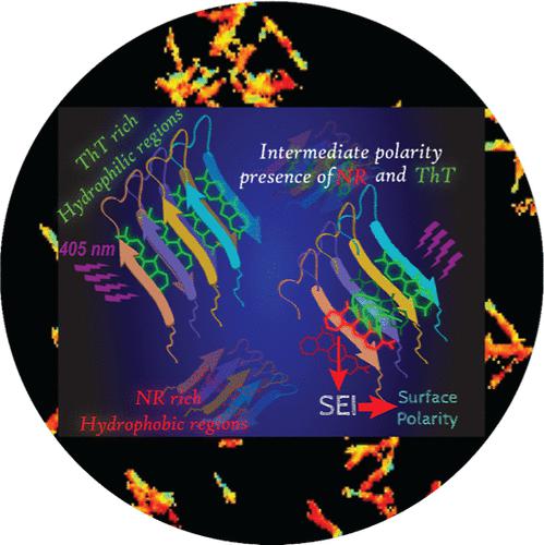
When misfolded, α-Synuclein (α-Syn), a natively disordered protein, aggregates to form amyloid fibrils responsible for the neurodegeneration observed in Parkinson’s disease. Structural studies revealed distinct molecular packing of α-Syn in different fibril polymorphs and variations of interprotofilament connections in the fibrillar architecture. Fibril polymorphs have been hypothesized to exhibit diverse surface polarities depending on the folding state of the protein during aggregation; however, the spatial variation of surface polarity in amyloid fibrils remains unexplored. To map the local polarity (or hydrophobicity) along α-Syn fibrils, we visualized the spectral characteristics of two dyes with distinct polarities–hydrophilic Thioflavin T (ThT) and hydrophobic Nile red (NR)─when both are bound to α-Syn fibrils. Dual-channel fluorescence imaging reveals uneven partitioning of ThT and NR along individual fibrils, implying that relatively more polar/hydrophobic patches are spread over a few hundred nanometers. Remarkably, spectrally resolved sensitized emission imaging of α-Syn fibrils provides unambiguous evidence of energy transfer from ThT to NR, implying that dyes of dissimilar polarity are in close proximity. Furthermore, spatially resolved fluorescence spectroscopy of the solvatochromic probe NR allowed us to quantitatively map the range and variation of the polarity parameter ET30 along individual fibrils. Our results suggest the existence of interlaced polar and nonpolar nanoscale domains throughout the fibrils; however, the relative populations of these patches vary considerably over larger length scales likely due to heterogeneous packing of α-Syn during fibrilization and dissimilar exposed polarities of polymorphic segments. The employed method may provide a foundation for imaging modalities of other similar structurally unresolved systems with diverse hydrophobic–hydrophilic topology.
期刊介绍:
ACS Chemical Neuroscience publishes high-quality research articles and reviews that showcase chemical, quantitative biological, biophysical and bioengineering approaches to the understanding of the nervous system and to the development of new treatments for neurological disorders. Research in the journal focuses on aspects of chemical neurobiology and bio-neurochemistry such as the following:
Neurotransmitters and receptors
Neuropharmaceuticals and therapeutics
Neural development—Plasticity, and degeneration
Chemical, physical, and computational methods in neuroscience
Neuronal diseases—basis, detection, and treatment
Mechanism of aging, learning, memory and behavior
Pain and sensory processing
Neurotoxins
Neuroscience-inspired bioengineering
Development of methods in chemical neurobiology
Neuroimaging agents and technologies
Animal models for central nervous system diseases
Behavioral research

 求助内容:
求助内容: 应助结果提醒方式:


